Enzyme classes:
General information:
|
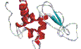
EC 3.2.1.17 - peptidoglycan N - acetylmuramoylhydrolase (lysozyme)
3D structures of EC 3.2.1.17 - lysozyme in Protein Data Bank
updated: 6 January 2022, 2:15
In total: 2051 PDB structures of EC 3.2.1.17 - lysozyme:
- 107l: Structural Basis of Alpha-helix Propensity at Two Sites in T4 Lysozyme
- 108l: Structural Basis of Alpha-helix Propensity at Two Sites in T4 Lysozyme
- 109l: Structural Basis of Alpha-helix Propensity at Two Sites in T4 Lysozyme
- 110l: Structural Basis of Alpha-helix Propensity at Two Sites in T4 Lysozyme
- 111l: Structural Basis of Alpha-helix Propensity at Two Sites in T4 Lysozyme
- 112l: Structural Basis of Alpha-helix Propensity at Two Sites in T4 Lysozyme
- 113l: Structural Basis of Alpha-helix Propensity at Two Sites in T4 Lysozyme
- 114l: Structural Basis of Alpha-helix Propensity at Two Sites in T4 Lysozyme
- 115l: Structural Basis of Alpha-helix Propensity at Two Sites in T4 Lysozyme
- 118l: The Energetic Cost and The Structural Consequences of Burying a Hydroxyl Group within The Core of a Protein Determined from Ala to Ser and Val to Thr Substitutions in T4 Lysozyme
- 119l: The Energetic Cost and The Structural Consequences of Burying a Hydroxyl Group within The Core of a Protein Determined from Ala to Ser and Val to Thr Substitutions in T4 Lysozyme
- 120l: The Energetic Cost and The Structural Consequences of Burying a Hydroxyl Group within The Core of a Protein Determined from Ala to Ser and Val to Thr Substitutions in T4 Lysozyme
- 122l: The Energetic Cost and The Structural Consequences of Burying a Hydroxyl Group within The Core of a Protein Determined from Ala to Ser and Val to Thr Substitutions in T4 Lysozyme
- 123l: The Energetic Cost and The Structural Consequences of Burying a Hydroxyl Group within The Core of a Protein Determined from Ala to Ser and Val to Thr Substitutions in T4 Lysozyme
- 125l: The Energetic Cost and The Structural Consequences of Burying a Hydroxyl Group within The Core of a Protein Determined from Ala to Ser and Val to Thr Substitutions in T4 Lysozyme
- 126l: The Energetic Cost and The Structural Consequences of Burying a Hydroxyl Group within The Core of a Protein Determined from Ala to Ser and Val to Thr Substitutions in T4 Lysozyme
- 127l: The Energetic Cost and The Structural Consequences of Burying a Hydroxyl Group within The Core of a Protein Determined from Ala to Ser and Val to Thr Substitutions in T4 Lysozyme
- 128l: The Energetic Cost and The Structural Consequences of Burying a Hydroxyl Group within The Core of a Protein Determined from Ala to Ser and Val to Thr Substitutions in T4 Lysozyme
- 129l: Structures of Randomly Generated Mutants of T4 Lysozyme Show That Protein Stability Can Be Enhanced by Relaxation of Strain and by Improved Hydrogen Bonding via Bound Solvent
- 130l: Structures of Randomly Generated Mutants of T4 Lysozyme Show That Protein Stability Can Be Enhanced by Relaxation of Strain and by Improved Hydrogen Bonding via Bound Solvent
- 131l: Structures of Randomly Generated Mutants of T4 Lysozyme Show That Protein Stability Can Be Enhanced by Relaxation of Strain and by Improved Hydrogen Bonding via Bound Solvent
- 132l: Structural Consequences of Reductive Methylation of Lysine Residues in Hen Egg White Lysozyme: an X-ray Analysis at 1.8 Angstroms Resolution
- 133l: Role of Arg 115 in The Catalytic Action of Human Lysozyme. X-ray Structure of His 115 and Glu 115 Mutants
- 134l: Role of Arg 115 in The Catalytic Action of Human Lysozyme. X-ray Structure of His 115 and Glu 115 Mutants
- 135l: X-ray Structure of Monoclinic Turkey Egg Lysozyme at 1.3 Angstroms Resolution
- 137l: Structural Basis of Amino Acid Alpha Helix Propensity
- 138l: Rapid Crystallization of T4 Lysozyme by Intermolecular Disulfide Crosslinking
- 139l: Rapid Crystallization of T4 Lysozyme by Intermolecular Disulfide Crosslinking
- 140l: Role of Backbone Flexibility in The Accommodation of Variants That Repack The Core of T4 Lysozyme
- 141l: Role of Backbone Flexibility in The Accommodation of Variants That Repack The Core of T4 Lysozyme
- 142l: Role of Backbone Flexibility in The Accommodation of Variants That Repack The Core of T4 Lysozyme
- 143l: Role of Backbone Flexibility in The Accommodation of Variants That Repack The Core of T4 Lysozyme
- 144l: Role of Backbone Flexibility in The Accommodation of Variants That Repack The Core of T4 Lysozyme
- 145l: Role of Backbone Flexibility in The Accommodation of Variants That Repack The Core of T4 Lysozyme
- 146l: Role of Backbone Flexibility in The Accommodation of Variants That Repack The Core of T4 Lysozyme
- 147l: Role of Backbone Flexibility in The Accommodation of Variants That Repack The Core of T4 Lysozyme
- 148l: A Covalent Enzyme-substrate Intermediate with Saccharide Distortion in a Mutant T4 Lysozyme
- 149l: Conservation of Solvent-binding Sites in 10 Crystal Forms of T4 Lysozyme
- 150l: Conservation of Solvent-binding Sites in 10 Crystal Forms of T4 Lysozyme
- 151l: Conservation of Solvent-binding Sites in 10 Crystal Forms of T4 Lysozyme
- 152l: Conservation of Solvent-binding Sites in 10 Crystal Forms of T4 Lysozyme
- 153l: The Refined Structures of Goose Lysozyme and Its Complex with a Bound Trisaccharide Show That The "goose-type Lysozymes Lack a Catalytic Aspartate
- 154l: The Refined Structures of Goose Lysozyme and Its Complex with a Bound Trisaccharide Show That The "goose-type Lysozymes Lack a Catalytic Aspartate
- 155l: Control of Enzyme Activity by an Engineered Disulfide Bond
- 156l: Control of Enzyme Activity by an Engineered Disulfide Bond
- 157l: Control of Enzyme Activity by an Engineered Disulfide Bond
- 158l: Control of Enzyme Activity by an Engineered Disulfide Bond
- 159l: Control of Enzyme Activity by an Engineered Disulfide Bond
- 160l: Control of Enzyme Activity by an Engineered Disulfide Bond
- 161l: Control of Enzyme Activity by an Engineered Disulfide Bond
- 162l: Control of Enzyme Activity by an Engineered Disulfide Bond
- 163l: Control of Enzyme Activity by an Engineered Disulfide Bond
- 164l: Control of Enzyme Activity by an Engineered Disulfide Bond
- 165l: Control of Enzyme Activity by an Engineered Disulfide Bond
- 166l: Control of Enzyme Activity by an Engineered Disulfide Bond
- 167l: Protein Flexibility and Adaptability Seen in 25 Crystal Forms of T4 Lysozyme
- 168l: Protein Flexibility and Adaptability Seen in 25 Crystal Forms of T4 Lysozyme
- 169l: Protein Flexibility and Adaptability Seen in 25 Crystal Forms of T4 Lysozyme
- 170l: Protein Flexibility and Adaptability Seen in 25 Crystal Forms of T4 Lysozyme
- 171l: Protein Flexibility and Adaptability Seen in 25 Crystal Forms of T4 Lysozyme
- 172l: Protein Flexibility and Adaptability Seen in 25 Crystal Forms of T4 Lysozyme
- 173l: Protein Flexibility and Adaptability Seen in 25 Crystal Forms of T4 Lysozyme
- 174l: Protein Flexibility and Adaptability Seen in 25 Crystal Forms of T4 Lysozyme
- 175l: Protein Flexibility and Adaptability Seen in 25 Crystal Forms of T4 Lysozyme
- 176l: Protein Flexibility and Adaptability Seen in 25 Crystal Forms of T4 Lysozyme
- 177l: Protein Flexibility and Adaptability Seen in 25 Crystal Forms of T4 Lysozyme
- 178l: Protein Flexibility and Adaptability Seen in 25 Crystal Forms of T4 Lysozyme
- 180l: Protein Flexibility and Adaptability Seen in 25 Crystal Forms of T4 Lysozyme
- 181l: Specificity of Ligand Binding in a Buried Non-polar Cavity of T4 Lysozyme: Linkage of Dynamics and Structural Plasticity
- 182l: Specificity of Ligand Binding in a Buried Non-polar Cavity of T4 Lysozyme: Linkage of Dynamics and Structural Plasticity
- 183l: Specificity of Ligand Binding in a Buried Non-polar Cavity of T4 Lysozyme: Linkage of Dynamics and Structural Plasticity
- 184l: Specificity of Ligand Binding in a Buried Non-polar Cavity of T4 Lysozyme: Linkage of Dynamics and Structural Plasticity
- 185l: Specificity of Ligand Binding in a Buried Non-polar Cavity of T4 Lysozyme: Linkage of Dynamics and Structural Plasticity
- 186l: Specificity of Ligand Binding in a Buried Non-polar Cavity of T4 Lysozyme: Linkage of Dynamics and Structural Plasticity
- 187l: Specificity of Ligand Binding in a Buried Non-polar Cavity of T4 Lysozyme: Linkage of Dynamics and Structural Plasticity
- 188l: Specificity of Ligand Binding in a Buried Non-polar Cavity of T4 Lysozyme: Linkage of Dynamics and Structural Plasticity
- 189l: Enhancement of Protein Stability by The Combination of Point Mutations in T4 Lysozyme Is Additive
- 190l: A Helix Initiation Signal in T4 Lysozyme Identified by Polyalanine Mutagenesis
- 191l: A Helix Initiation Signal in T4 Lysozyme Identified by Polyalanine Mutagenesis
- 192l: A Helix Initiation Signal in T4 Lysozyme Identified by Polyalanine Mutagenesis
- 195l: Thermodynamic and Structural Compensation in "size-switch" Core-repacking Variants of T4 Lysozyme
- 196l: Thermodynamic and Structural Compensation in "size-switch" Core-repacking Variants of T4 Lysozyme
- 197l: Thermodynamic and Structural Compensation in "size-switch" Core-repacking Variants of T4 Lysozyme
- 198l: Thermodynamic and Structural Compensation in "size-switch" Core-repacking Variants of T4 Lysozyme
- 199l: Thermodynamic and Structural Compensation in "size-switch" Core-repacking Variants of T4 Lysozyme
- 1a2y: Hen Egg White Lysozyme, D18A Mutant, in Complex with Mouse Monoclonal Antibody D1.3
- 1aki: The Structure of The Orthorhombic Form of Hen Egg-white Lysozyme at 1.5 Angstroms Resolution
- 1am7: Lysozyme from Bacteriophage Lambda
- 1at5: Hen Egg White Lysozyme with a Succinimide Residue
- 1at6: Hen Egg White Lysozyme with a Isoaspartate Residue
- 1azf: Chicken Egg White Lysozyme Crystal Grown in Bromide Solution
- 1b0d: Structural Effects of Monovalent Anions on Polymorphic Lysozyme Crystals
- 1b2k: Structural Effects of Monovalent Anions on Polymorphic Lysozyme Crystals
- 1b5u: Contribution of Hydrogen Bonds to The Conformational Stability of Human Lysozyme: Calorimetry and X-ray Analysis of Six Ser->ala Mutant
- 1b5v: Contribution of Hydrogen Bonds to The Conformational Stability of Human Lysozyme: Calorimetry and X-ray Analysis of Six Ser->ala Mutants
- 1b5w: Contribution of Hydrogen Bonds to The Conformational Stability of Human Lysozyme: Calorimetry and X-ray Analysis of Six Ser->ala Mutants
- 1b5x: Contribution of Hydrogen Bonds to The Conformational Stability of Human Lysozyme: Calorimetry and X-ray Analysis of Six Ser->ala Mutants
- 1b5y: Contribution of Hydrogen Bonds to The Conformational Stability of Human Lysozyme: Calorimetry and X-ray Analysis of Six Ser->ala Mutants
- 1b5z: Contribution of Hydrogen Bonds to The Conformational Stability of Human Lysozyme: Calorimetry and X-ray Analysis of Six Ser->ala Mutants
- 1b6i: T4 Lysozyme Mutant with Cys 54 Replaced by Thr, Cys 97 Replaced by Ala, Thr 21 Replaced by Cys and Lys 124 Replaced by Cys (C54T,C97A,T21C,K124C)
- 1b7l: Verification of Spmp Using Mutant Human Lysozymes
- 1b7m: Verification of Spmp Using Mutant Human Lysozymes
- 1b7n: Verification of Spmp Using Mutant Human Lysozymes
- 1b7o: Verification of Spmp Using Mutant Human Lysozymes
- 1b7p: Verification of Spmp Using Mutant Human Lysozymes
- 1b7q: Verification of Spmp Using Mutant Human Lysozymes
- 1b7r: Verification of Spmp Using Mutant Human Lysozymes
- 1bb3: Human Lysozyme Mutant A96L
- 1bb4: Human Lysozyme Double Mutant A96L, W109H
- 1bb5: Human Lysozyme Mutant A96L Complexed with Chitotriose
- 1bb6: Lysozyme Complex with 4-methyl-umbelliferyl Chitotriose
- 1bb7: Lysozyme Complex with 4-methyl-umbelliferyl Chitobiose
- 1bgi: Orthorhombic Lysozyme Crystallized at High Temperature (310k)
- 1bhz: Low Temperature Middle Resolution Structure of Hen Egg White Lysozyme from Masc Data
- 1bvk: Humanized Anti-lysozyme Fv Complexed with Lysozyme
- 1bvx: The 1.8 a Structure of Gel Grown Tetragonal Hen Egg White Lysozyme
- 1bwh: The 1.8 a Structure of Ground Control Grown Tetragonal Hen Egg White Lysozyme
- 1bwi: The 1.8 a Structure of Microbatch Oil Drop Grown Tetragonal Hen Egg White Lysozyme
- 1bwj: The 1.8 a Structure of Microgravity Grown Tetragonal Hen Egg White Lysozyme
- 1c10: Crystal Structure of Hew Lysozyme under Pressure of Xenon (8 Bar)
- 1c43: Mutant Human Lysozyme with Foreign N-terminal Residues
- 1c45: Mutant Human Lysozyme with Foreign N-terminal Residues
- 1c46: Mutant Human Lysozyme with Foreign N-terminal Residues
- 1c60: T4 Lysozyme Mutant C54T/C97A/F153A in The Presence of 8 Atm Argon
- 1c61: T4 Lysozyme Mutant C54T/C97A/F153A in The Presence of 8 Atm Krypton
- 1c62: T4 Lysozyme Mutant C54T/C97A/F153A in The Presence of 8 Atm Xenon
- 1c63: T4 Lysozyme Mutant C54T/C97A/L121A in The Presence of 8 Atm Argon
- 1c64: T4 Lysozyme Mutant C54T/C97A/L121A in The Presence of 8 Atm Krypton
- 1c65: T4 Lysozyme Mutant C54T/C97A/L121A in The Presence of 8 Atm Xenon
- 1c66: T4 Lysozyme Mutant C54T/C97A/L121A/L133A in The Presence of 8 Atm Argon
- 1c67: T4 Lysozyme Mutant C54T/C97A/L121A/L133A in The Presence of 8 Atm Krypton
- 1c68: T4 Lysozyme Mutant C54T/C97A/L121A/L133A in The Presence of 8 Atm Xenon
- 1c69: T4 Lysozyme Mutant C54T/C97A/L133A in The Presence of 8 Atm Argon
- 1c6a: T4 Lysozyme Mutant C54T/C97A/L133A in The Presence of 8 Atm Krypton
- 1c6b: T4 Lysozyme Mutant C54T/C97A/L133A in The Presence of 8 Atm Xenon
- 1c6c: T4 Lysozyme Mutant C54T/C97A/L99A in The Presence of 16 Atm Argon
- 1c6d: T4 Lysozyme Mutant C54T/C97A/L99A in The Presence of 16 Atm Krypton
- 1c6e: T4 Lysozyme Mutant C54T/C97A/L99A in The Presence of 2 Atm Xenon
- 1c6f: T4 Lysozyme Mutant C54T/C97A/L99A in The Presence of 32 Atm Argon
- 1c6g: T4 Lysozyme Mutant C54T/C97A/L99A in The Presence of 4 Atm Krypton
- 1c6h: T4 Lysozyme Mutant C54T/C97A/L99A in The Presence of 4 Atm Xenon
- 1c6i: T4 Lysozyme Mutant C54T/C97A/L99A in The Presence of 8 Atm Argon
- 1c6j: T4 Lysozyme Mutant C54T/C97A/L99A in The Presence of 8 Atm Krypton
- 1c6k: T4 Lysozyme Mutant C54T/C97A/L99A in The Presence of 8 Atm Xenon
- 1c6l: T4 Lysozyme Mutant C54T/C97A/L99A/F153A in The Presence of 8 Atm Argon
- 1c6m: T4 Lysozyme Mutant C54T/C97A/L99A/F153A in The Presence of 8 Atm Krypton
- 1c6n: T4 Lysozyme Mutant C54T/C97A/L99A/F153A in The Presence of 8 Atm Xenon
- 1c6p: T4 Lysozyme Mutant C54T/C97A in The Presence of 8 Atm Argon
- 1c6q: T4 Lysozyme Mutant C54T/C97A in The Presence of 8 Atm Krypton
- 1c6t: T4 Lysozyme Mutant C54T/C97A in The Presence of 8 Atm Xenon
- 1c7p: Crystal Structure of Mutant Human Lysozyme with Four Extra Residues (eaea) at The N-terminal
- 1cj6: T11A Mutant Human Lysozyme
- 1cj7: T11V Mutant Human Lysozyme
- 1cj8: T40A Mutant Human Lysozyme
- 1cj9: T40V Mutant Human Lysozyme
- 1ckc: T43A Mutant Human Lysozyme
- 1ckd: T43V Mutant Human Lysozyme
- 1ckf: T52A Mutant Human Lysozyme
- 1ckg: T52V Mutant Human Lysozyme
- 1ckh: T70V Mutant Human Lysozyme
- 1ctw: T4 Lysozyme Mutant I78A
- 1cu0: T4 Lysozyme Mutant I78M
- 1cu2: T4 Lysozyme Mutant L84M
- 1cu3: T4 Lysozyme Mutant V87M
- 1cu5: T4 Lysozyme Mutant L91M
- 1cu6: T4 Lysozyme Mutant L91A
- 1cup: Methionine Core Mutant of T4 Lysozyme
- 1cuq: T4 Lysozyme Mutant V103M
- 1cv0: T4 Lysozyme Mutant F104M
- 1cv1: T4 Lysozyme Mutant V111M
- 1cv3: T4 Lysozyme Mutant L121M
- 1cv4: T4 Lysozyme Mutant L118M
- 1cv5: T4 Lysozyme Mutant L133M
- 1cv6: T4 Lysozyme Mutant V149M
- 1cvk: T4 Lysozyme Mutant L118A
- 1cx6: T4 Lysozyme Substituted with Selenomethionine
- 1cx7: T4 Lysozyme Methionine Core Mutant
- 1d2w: N-terminal Domain Core Methionine Mutation
- 1d2y: N-terminal Domain Core Methionine Mutation
- 1d3f: N-terminal Domain Core Methionine Mutation
- 1d3j: N-terminal Domain Core Methionine Mutation
- 1d3m: Methionine Core Mutation
- 1d3n: Methionine Core Mutation
- 1d6p: Human Lysozyme L63 Mutant Labelled with 2',3'-epoxypropyl N, N'-diacetylchitobiose
- 1d6q: Human Lysozyme E102 Mutant Labelled with 2',3'-epoxypropyl Glycoside of N-acetyllactosamine
- 1d9u: Bacteriophage Lambda Lysozyme Complexed with a Chitohexasacharide
- 1d9w: Bacteriophage T4 Lysozyme Mutant
- 1di3: Role of Amino Acid Residues at Turns in The Conformational Stability and Folding of Human Lysozyme
- 1di4: Role of Amino Acid Residues at Turns in The Conformational Stability and Folding of Human Lysozyme
- 1di5: Role of Amino Acid Residues at Turns in The Conformational Stability and Folding of Human Lysozyme
- 1dkj: Bobwhite Quail Lysozyme
- 1dkk: Bobwhite Quail Lysozyme with Nitrate
- 1dpw: Structure of Hen Egg-white Lysozyme in Complex with Mpd
- 1dpx: Structure of Hen Egg-white Lysozyme
- 1dqj: Crystal Structure of The Anti-lysozyme Antibody Hyhel-63 Complexed with Hen Egg White Lysozyme
- 1dya: Determination of Alpha-helix Propensity within The Context of a Folded Protein: Sites 44 and 131 in Bacteriophage T4 Lysozyme
- 1dyb: Determination of Alpha-helix Propensity within The Context of a Folded Protein: Sites 44 and 131 in Bacteriophage T4 Lysozyme
- 1dyc: Determination of Alpha-helix Propensity within The Context of a Folded Protein: Sites 44 and 131 in Bacteriophage T4 Lysozyme
- 1dyd: Determination of Alpha-helix Propensity within The Context of a Folded Protein: Sites 44 and 131 in Bacteriophage T4 Lysozyme
- 1dye: Determination of Alpha-helix Propensity within The Context of a Folded Protein: Sites 44 and 131 in Bacteriophage T4 Lysozyme
- 1dyf: Determination of Alpha-helix Propensity within The Context of a Folded Protein: Sites 44 and 131 in Bacteriophage T4 Lysozyme
- 1dyg: Determination of Alpha-helix Propensity within The Context of a Folded Protein: Sites 44 and 131 in Bacteriophage T4 Lysozyme
- 1dzb: Crystal Structure of Phage Library-derived Single-chain Fv Fragment 1f9 in Complex with Turkey Egg-white Lysozyme
- 1e8l: Nmr Solution Structure of Hen Lysozyme
- 1el1: X-ray Crystal Structure Analysis of Canine Milk Lysozyme (holo-type)
- 1epy: T4 Lysozyme Mutant, T21H/C54T/C97A/Q141H/T142H
- 1eq4: Crystal Structures of Salt Bridge Mutants of Human Lysozyme
- 1eq5: Crystal Structures of Salt Bridge Mutants of Human Lysozyme
- 1eqe: Crystal Structures of Salt Bridge Mutants of Human Lysozyme
- 1f0w: Crystal Structure of Orthorhombic Lysozyme Grown at Ph 6.5
- 1f10: Crystal Structure of Orthorhombic Lysozyme Grown at Ph 6.5 at 88% Relative Humidity
- 1fbi: Crystal Structure of a Cross-reaction Complex between Fab F9.13.7 and Guinea-fowl Lysozyme
- 1fdl: Crystallographic Refinement of The Three-dimensional Structure of The Fab D1.3-lysozyme Complex at 2.5- Angstroms Resolution
- 1flq: Hen Egg White Lysozyme Mutant with Alanine Substituted for Glycine
- 1flu: Hen Egg White Lysozyme Mutant with Alanine Substituted for Glycine
- 1flw: Hen Egg White Lysozyme Mutant with Alanine Substituted for Glycine
- 1fly: Hen Egg White Lysozyme Mutant with Alanine Substituted for Glycine
- 1fn5: Hen Egg White Lysozyme Mutant with Alanine Substituted for Glycine
- 1g06: Crystal Structure of T4 Lysozyme Mutant V149S
- 1g07: Crystal Structure of T4 Lysozyme Mutant V149C
- 1g0g: Crystal Structure of T4 Lysozyme Mutant T152A
- 1g0j: Crystal Structure of T4 Lysozyme Mutant T152S
- 1g0k: Crystal Structure of T4 Lysozyme Mutant T152C
- 1g0l: Crystal Structure of T4 Lysozyme Mutant T152V
- 1g0m: Crystal Structure of T4 Lysozyme Mutant T152I
- 1g0p: Crystal Structure of T4 Lysozyme Mutant V149G
- 1g0q: Crystal Structure of T4 Lysozyme Mutant V149I
- 1g1v: T4 Lysozyme Mutant C54T/C97A/I58T
- 1g1w: T4 Lysozyme Mutant C54T/C97A/Q105M
- 1g7h: Crystal Structure of Hen Egg White Lysozyme (hel) Complexed with The Mutant Anti-hel Monoclonal Antibody D1.3(VLW92A)
- 1g7i: Crystal Structure of Hen Egg White Lysozyme (hel) Complexed with The Mutant Anti-hel Monoclonal Antibody D1.3 (VLW92F)
- 1g7j: Crystal Structure of Hen Egg White Lysozyme (hel) Complexed with The Mutant Anti-hel Monoclonal Antibody D1.3 (VLW92H)
- 1g7l: Crystal Structure of Hen Egg White Lysozyme (hel) Complexed with The Mutant Anti-hel Monoclonal Antibody D1.3 (VLW92S)
- 1g7m: Crystal Structure of Hen Egg White Lysozyme (hel) Complexed with The Mutant Anti-hel Monoclonal Antibody D1.3 (VLW92V)
- 1gay: Crystal Structure of Mutant Human Lysozyme Substituted at The Surface Positions
- 1gaz: Crystal Structure of Mutant Human Lysozyme Substituted at The Surface Positions
- 1gb0: Crystal Structure of Mutant Human Lysozyme Substituted at The Surface Positions
- 1gb2: Crystal Structure of Mutant Human Lysozyme Substituted at The Surface Positions
- 1gb3: Crystal Structure of Mutant Human Lysozyme Substituted at The Surface Positions
- 1gb5: Crystal Structure of Mutant Human Lysozyme Substituted at The Surface Positions
- 1gb6: Crystal Structure of Mutant Human Lysozyme Substituted at The Surface Positions
- 1gb7: Crystal Structure of Mutant Human Lysozyme Substituted at The Surface Positions
- 1gb8: Crystal Structure of Mutant Human Lysozyme Substituted at The Surface Positions
- 1gb9: Crystal Structure of Mutant Human Lysozyme Substituted at The Surface Positions
- 1gbo: Crystal Structure of Mutant Human Lysozyme Substituted at The Surface Positions
- 1gbs: Crystal Structure of Black Swan Goose-type Lysozyme at 1.8 Angstroms Resolution
- 1gbw: Crystal Structure of Mutant Human Lysozyme Substituted at The Surface Positions
- 1gbx: Crystal Structure of Mutant Human Lysozyme Substituted at The Surface Positions
- 1gby: Crystal Structure of Mutant Human Lysozyme Substituted at The Surface Positions
- 1gbz: Crystal Structure of Mutant Human Lysozyme Substituted at The Surface Positions
- 1gd6: Structure of The Bombyx Mori Lysozyme
- 1gdw: Crystal Structure of Mutant Human Lysozyme Substituted at Left-handed Helical Positions
- 1gdx: Crystal Structure of Mutant Human Lysozyme Substituted at Left-handed Helical Positions
- 1ge0: Crystal Structure of Mutant Human Lysozyme Substituted at Left-handed Helical Positions
- 1ge1: Crystal Structure of Mutant Human Lysozyme Substituted at Left-handed Helical Positions
- 1ge2: Crystal Structure of Mutant Human Lysozyme Substituted at Left-handed Helical Positions
- 1ge3: Crystal Structure of Mutant Human Lysozyme Substituted at Left-handed Helical Positions
- 1ge4: Crystal Structure of Mutant Human Lysozyme Substituted at Left-handed Helical Positions
- 1gev: Buried Polar Mutant Human Lysozyme
- 1gez: Buried Polar Mutant Human Lysozyme
- 1gf0: Buried Polar Mutant Human Lysozyme
- 1gf3: Buried Polar Mutant Human Lysozyme
- 1gf4: Buried Polar Mutant Human Lysozyme
- 1gf5: Buried Polar Mutant Human Lysozyme
- 1gf6: Buried Polar Mutant Human Lysozyme
- 1gf7: Buried Polar Mutant Human Lysozyme
- 1gf8: Crystal Structure of Mutant Human Lysozyme Substituted at The Surface Positions
- 1gf9: Crystal Structure of Mutant Human Lysozyme Substituted at The Surface Positions
- 1gfa: Crystal Structure of Mutant Human Lysozyme Substituted at The Surface Positions
- 1gfe: Crystal Structure of Mutant Human Lysozyme Substituted at The Surface Positions
- 1gfg: Crystal Structure of Mutant Human Lysozyme Substituted at The Surface Positions
- 1gfh: Crystal Structure of Mutant Human Lysozyme Substituted at The Surface Positions
- 1gfj: Crystal Structure of Mutant Human Lysozyme Substituted at The Surface Positions
- 1gfk: Crystal Structure of Mutant Human Lysozyme Substituted at The Surface Positions
- 1gfr: Crystal Structure of Mutant Human Lysozyme Substituted at The Surface Positions
- 1gft: Crystal Structure of Mutant Human Lysozyme Substituted at The Surface Positions
- 1gfu: Crystal Structure of Mutant Human Lysozyme Substituted at The Surface Positions
- 1gfv: Crystal Structure of Mutant Human Lysozyme Substituted at The Surface Positions
- 1ghl: The Three-dimensional Structure of Pheasant and Guinea-fowl Egg Lysozymes
- 1gpq: Structure of Ivy Complexed with Its Target, Hewl
- 1gwd: Tri-iodide Derivative of Hen Egg-white Lysozyme
- 1gxv: Solution Structure of Lysozyme at Low and High Pressure
- 1gxx: Solution Structure of Lysozyme at Low and High Pressure
- 1h09: Multimodular Pneumococcal Cell Wall Endolysin from Phage Cp-1
- 1h6m: Covalent Glycosyl-enzyme Intermediate of Hen Egg White Lysozyme
- 1h87: Gadolinium Derivative of Tetragonal Hen Egg-white Lysozyme at 1.7 a Resolution
- 1hc0: Structure of Lysozyme with Periodate
- 1hel: Structural and Thermodynamic Analysis of Compensating Mutations within The Core of Chicken Egg White Lysozyme
- 1hem: Structural and Thermodynamic Analysis of Compensating Mutations within The Core of Chicken Egg White Lysozyme
- 1hen: Structural and Thermodynamic Analysis of Compensating Mutations within The Core of Chicken Egg White Lysozyme
- 1heo: Structural and Thermodynamic Analysis of Compensating Mutations within The Core of Chicken Egg White Lysozyme
- 1hep: Structural and Thermodynamic Analysis of Compensating Mutations within The Core of Chicken Egg White Lysozyme
- 1heq: Structural and Thermodynamic Analysis of Compensating Mutations within The Core of Chicken Egg White Lysozyme
- 1her: Structural and Thermodynamic Analysis of Compensating Mutations within The Core of Chicken Egg White Lysozyme
- 1hew: Refinement of an Enzyme Complex with Inhibitor Bound at Partial Occupancy. Hen Egg-white Lysozyme and Tri-n- Acetylchitotriose at 1.75 Angstroms Resolution
- 1hf4: Structural Effects of Monovalent Anions on Polymorphic Lysozyme Crystals
- 1hhl: The Three-dimensional Structure of Pheasant and Guinea-fowl Egg Lysozymes
- 1hnl: Crystal Structure of a Glutathionylated Human Lysozyme: a Folding Intermediate Mimic in The Formation of a Disulfide Bond
- 1hsw: Lysozyme (mucopeptide N-acetylmuramyl Hydrolase)
- 1hsx: Lysozyme Grown at Basic Ph and Its Low Humidity Variant
- 1hvq: Crystal Structures of Hevamine, a Plant Defence Protein with Chitinase and Lysozyme Activity, and Its Complex with an Inhibitor
- 1i22: Mutant Human Lysozyme (A83K/Q86D/A92D)
- 1i56: Solution Structure of Ca2+-bound State of Canine Milk Lysozyme
- 1i6s: T4 Lysozyme Mutant C54T/C97A/N101A
- 1ic4: Crystal Structure of Hyhel-10 Fv MUTANT(HD32A)-HEN Lysozyme Complex
- 1ic5: Crystal Structure of Hyhel-10 Fv MUTANT(HD99A)-HEN Lysozyme Complex
- 1ic7: Crystal Structure of Hyhel-10 Fv MUTANT(HD32A99A)-HEN Lysozyme Complex
- 1iee: Structure of Tetragonal Hen Egg White Lysozyme at 0.94 a from Crystals Grown by The Counter-diffusion Method
- 1iiz: Crystal Structure of The Induced Antibacterial Protein from Tasar Silkworm, Antheraea Mylitta
- 1inu: Crystal Structure of Mutant Human Lysozyme Substituted at The Surface Positions
- 1io5: Hydrogen and Hydration of Hen Egg-white Lysozyme Determined by Neutron Diffraction
- 1ioc: Crystal Structure of Mutant Human Lysozyme, EAEA-I56T
- 1ioq: Stabilization of Hen Egg White Lysozyme by a Cavity-filling Mutation
- 1ior: Stabilization of Hen Egg White Lysozyme by a Cavity-filling Mutation
- 1ios: Stabilization of Hen Egg White Lysozyme by a Cavity-filling Mutation
- 1ip1: G37A Human Lysozyme
- 1ip2: G48A Human Lysozyme
- 1ip3: G68A Human Lysozyme
- 1ip4: G72A Human Lysozyme
- 1ip5: G105A Human Lysozyme
- 1ip6: G127A Human Lysozyme
- 1ip7: G129A Human Lysozyme
- 1ir7: Im Mutant of Lysozyme
- 1ir8: Im Mutant of Lysozyme
- 1ir9: Im Mutant of Lysozyme
- 1ivm: Solution Structure of Mouse Lysozyme M
- 1iwt: Crystal Structure Analysis of Human Lysozyme at 113k.
- 1iwu: Crystal Structure Analysis of Human Lysozyme at 127k.
- 1iwv: Crystal Structure Analysis of Human Lysozyme at 147k.
- 1iww: Crystal Structure Analysis of Human Lysozyme at 152k.
- 1iwx: Crystal Structure Analysis of Human Lysozyme at 161k.
- 1iwy: Crystal Structure Analysis of Human Lysozyme at 170k.
- 1iwz: Crystal Structure Analysis of Human Lysozyme at 178k.
- 1ix0: I59A-3SS Human Lysozyme
- 1iy3: Solution Structure of The Human Lysozyme at 4 Degree C
- 1iy4: Solution Structure of The Human Lysozyme at 35 Degree C
- 1j1o: Crystal Structure of Hyhel-10 Fv Mutant LY50F Complexed with Hen Egg White Lysozyme
- 1j1p: Crystal Structure of Hyhel-10 Fv Mutant LS91A Complexed with Hen Egg White Lysozyme
- 1j1x: Crystal Structure of Hyhel-10 Fv Mutant LS93A Complexed with Hen Egg White Lysozyme
- 1ja2: Binding of N-acetylglucosamine to Chicken Egg Lysozyme: a Powder Diffraction Study
- 1ja4: Binding of N-acetylglucosamine to Chicken Egg Lysozyme: a Powder Diffraction Study
- 1ja6: Binding of N-acetylglucosamine to Chicken Egg Lysozyme: a Powder Diffraction Study
- 1ja7: Binding of N-acetylglucosamine to Chicken Egg Lysozyme: a Powder Diffraction Study
- 1jef: Turkey Lysozyme Complex with (glcnac)3
- 1jfx: Crystal Structure of The Bacterial Lysozyme from Streptomyces Coelicolor at 1.65 a Resolution
- 1jis: Crystal Structure of Tetragonal Lysozyme Grown at Ph 4.6
- 1jit: Crystal Structure of Tetragonal Lysozyme Grown in Presence 30% Trehalose
- 1jiy: Crystal Structure of Tetragonal Lysozyme Grown in Presence 20% Sorbitol
- 1jj0: Crystal Structure of Tetragonal Lysozyme Grown in Presence of 30% Sucrose
- 1jj1: Crystal Structure of Orthorhombic Lysozyme Grown at Ph 4.6 in Presence of 5% Sorbitol
- 1jj3: Crystal Structure of Monoclinic Lysozyme Grown at Ph 4.6
- 1jka: Human Lysozyme Mutant with Glu 35 Replaced by Asp
- 1jkb: Human Lysozyme Mutant with Glu 35 Replaced by Ala
- 1jkc: Human Lysozyme Mutant with Trp 109 Replaced by Phe
- 1jkd: Human Lysozyme Mutant with Trp 109 Replaced by Ala
- 1jpo: Low Temperature Orthorhombic Lysozyme
- 1jqu: Are Carboxy Terminii of Helices Coded by The Local Sequence or by Tertiary Structure Contacts
- 1jse: Full-matrix Least-squares Refinement of Turkey Lysozyme
- 1jsf: Full-matrix Least-squares Refinement of Human Lysozyme
- 1jtm: Alternative Structures of a Sequence Extended T4 Lysozyme Show That The Highly Conserved Beta-sheet Has Weak Intrinsic Folding Propensity
- 1jtn: Alternative Structures of a Sequence Extended T4 Lysozyme Show That The Highly Conserved Beta-sheet Region Has Weak Intrinsic Folding Propensity
- 1jto: Degenerate Interfaces in Antigen-antibody Complexes
- 1jtp: Degenerate Interfaces in Antigen-antibody Complexes
- 1jtt: Degenerate Interfaces in Antigen-antibody Complexes
- 1jug: Lysozyme from Echidna Milk (tachyglossus Aculeatus)
- 1jwr: Crystal Structure of Human Lysozyme at 100 K
- 1k28: The Structure of The Bacteriophage T4 Cell-puncturing Device
- 9lyz: X-ray Crystallography of The Binding of The Bacterial Cell Wall Trisaccharide Nam-nag-nam to Lysozyme
- 8lyz: An X-ray Study of The Structure and Binding Properties of Iodine-inactivated Lysozyme
- 7lzm: Comparison of The Crystal Structure of Bacteriophage T4 Lysozyme at Low, Medium, and High Ionic Strengths
- 7lyz: Protein Model Building by The Use of a Constrained- Restrained Least-squares Procedure
- 6lzm: Comparison of The Crystal Structure of Bacteriophage T4 Lysozyme at Low, Medium, and High Ionic Strengths
- 6lyz: Real-space Refinement of The Structure of Hen Egg-white Lysozyme
- 6lyt: Comparison of Radiation-induced Decay and Structure Refinement from X-ray Data Collected from Lysozyme Crystals at Low and Ambient Temperatures
- 5lzm: Comparison of The Crystal Structure of Bacteriophage T4 Lysozyme at Low, Medium, and High Ionic Strengths
- 5lyz: Real-space Refinement of The Structure of Hen Egg-white Lysozyme
- 5lyt: Comparison of Radiation-induced Decay and Structure Refinement from X-ray Data Collected from Lysozyme Crystals at Low and Ambient Temperatures
- 5lym: Studies of Monoclinic Hen Egg White Lysozyme. Iv. X-ray Refinement at 1.8 Angstrom Resolution and a Comparison of The Variable Regions in The Polymorphic Forms
- 4lzt: Atomic Resolution Refinement of Triclinic Hew Lysozyme at 295k
- 4lzm: Comparison of The Crystal Structure of Bacteriophage T4 Lysozyme at Low, Medium, and High Ionic Strengths
- 4lyz: Real-space Refinement of The Structure of Hen Egg-white Lysozyme
- 4lyt: Comparison of Radiation-induced Decay and Structure Refinement from X-ray Data Collected from Lysozyme Crystals at Low and Ambient Temperatures
- 4lyo: Cross-linked Chicken Lysozyme Crystal in Neat Acetonitrile, Then Back-soaked in Water
- 4lym: Crystal Structure of Low Humidity Tetragonal Lysozyme at 2.1-angstroms Resolution. Variability in Hydration Shell and Its Structural Consequences
- 1kip: Fv Mutant Y(b 32)a (vh Domain) of Mouse Monoclonal Antibody D1.3 Complexed with Hen Egg White Lysozyme
- 1kiq: Fv Mutant Y(b 101)f (vh Domain) of Mouse Monoclonal Antibody D1.3 Complexed with Hen Egg White Lysozyme
- 1kir: Fv Mutant Y(a 50)s (vl Domain) of Mouse Monoclonal Antibody D1.3 Complexed with Hen Egg White Lysozyme
- 1kni: Stabilizing Disulfide Bridge Mutant of T4 Lysozyme
- 3pbl: Structure of The Human Dopamine D3 Receptor in Complex with Eticlopride
- 1kqy: Hevamine Mutant D125A/E127A/Y183F in Complex with Penta-nag
- 1kqz: Hevamine Mutant D125A/E127A/Y183F in Complex with Tetra-nag
- 1kr0: Hevamine Mutant D125A/Y183F in Complex with Tetra-nag
- 1kr1: Hevamine Mutant D125A/E127A in Complex with Tetra-nag
- 1ks3: Methionine Core Mutant of T4 Lysozyme
- 1kw5: Methionine Core Mutant of T4 Lysozyme
- 1kw7: Methionine Core Mutant of T4 Lysozyme
- 1kxw: Analysis of The Stabilization of Hen Lysozyme with The Helix Dipole and Charged Side Chains
- 1kxx: Analysis of The Stabilization of Hen Lysozyme with The Helix Dipole and Charged Side Chains
- 1kxy: Analysis of The Stabilization of Hen Lysozyme with The Helix Dipole and Charged Side Chains
- 1ky0: Methionine Core Mutant of T4 Lysozyme
- 1ky1: Methionine Core Mutant of T4 Lysozyme
- 1l00: Perturbation of Trp 138 in T4 Lysozyme by Mutations at Gln 105 Used to Correlate Changes in Structure, Stability, Solvation, and Spectroscopic Properties
- 1l01: Structural Studies of Mutants of The Lysozyme of Bacteriophage T4. The Temperature-sensitive Mutant Protein Thr157 (right Arrow) Ile
- 1l02: Contributions of Hydrogen Bonds of Thr 157 to The Thermodynamic Stability of Phage T4 Lysozyme
- 1l03: Contributions of Hydrogen Bonds of Thr 157 to The Thermodynamic Stability of Phage T4 Lysozyme
- 1l04: Contributions of Hydrogen Bonds of Thr 157 to The Thermodynamic Stability of Phage T4 Lysozyme
- 1l05: Contributions of Hydrogen Bonds of Thr 157 to The Thermodynamic Stability of Phage T4 Lysozyme
- 1l06: Contributions of Hydrogen Bonds of Thr 157 to The Thermodynamic Stability of Phage T4 Lysozyme
- 1l07: Contributions of Hydrogen Bonds of Thr 157 to The Thermodynamic Stability of Phage T4 Lysozyme
- 1l08: Contributions of Hydrogen Bonds of Thr 157 to The Thermodynamic Stability of Phage T4 Lysozyme
- 1l09: Contributions of Hydrogen Bonds of Thr 157 to The Thermodynamic Stability of Phage T4 Lysozyme
- 1l0j: Methionine Core Mutant of T4 Lysozyme
- 1l0k: Methionine Core Mutant of T4 Lysozyme
- 1l10: Structural Studies of Mutants of The Lysozyme of Bacteriophage T4. The Temperature-sensitive Mutant Protein Thr157 (right Arrow) Ile
- 1l11: Contributions of Hydrogen Bonds of Thr 157 to The Thermodynamic Stability of Phage T4 Lysozyme
- 1l12: Contributions of Hydrogen Bonds of Thr 157 to The Thermodynamic Stability of Phage T4 Lysozyme
- 1l13: Contributions of Hydrogen Bonds of Thr 157 to The Thermodynamic Stability of Phage T4 Lysozyme
- 1l14: Contributions of Hydrogen Bonds of Thr 157 to The Thermodynamic Stability of Phage T4 Lysozyme
- 1l15: Contributions of Hydrogen Bonds of Thr 157 to The Thermodynamic Stability of Phage T4 Lysozyme
- 1l16: Structural Analysis of The Temperature-sensitive Mutant of Bacteriophage T4 Lysozyme, Glycine 156 (right Arrow) Aspartic Acid
- 1l17: Hydrophobic Stabilization in T4 Lysozyme Determined Directly by Multiple Substitutions of Ile 3
- 1l18: Hydrophobic Stabilization in T4 Lysozyme Determined Directly by Multiple Substitutions of Ile 3
- 1l19: Enhanced Protein Thermostability from Designed Mutations That Interact with Alpha-helix Dipoles
- 1l20: Enhanced Protein Thermostability from Designed Mutations That Interact with Alpha-helix Dipoles
- 1l21: Contributions of Left-handed Helical Residues to The Structure and Stability of Bacteriophage T4 Lysozyme
- 1l22: Contributions of Left-handed Helical Residues to The Structure and Stability of Bacteriophage T4 Lysozyme
- 1l23: Enhanced Protein Thermostability from Site-directed Mutations That Decrease The Entropy of Unfolding
- 1l24: Enhanced Protein Thermostability from Site-directed Mutations That Decrease The Entropy of Unfolding
- 1l25: Replacements of Pro86 in Phage T4 Lysozyme Extend an Alpha- Helix but Do Not Alter Protein Stability
- 1l26: Replacements of Pro86 in Phage T4 Lysozyme Extend an Alpha- Helix but Do Not Alter Protein Stability
- 1l27: Replacements of Pro86 in Phage T4 Lysozyme Extend an Alpha- Helix but Do Not Alter Protein Stability
- 1l28: Replacements of Pro86 in Phage T4 Lysozyme Extend an Alpha- Helix but Do Not Alter Protein Stability
- 1l29: Replacements of Pro86 in Phage T4 Lysozyme Extend an Alpha- Helix but Do Not Alter Protein Stability
- 1l30: Replacements of Pro86 in Phage T4 Lysozyme Extend an Alpha- Helix but Do Not Alter Protein Stability
- 1l31: Replacements of Pro86 in Phage T4 Lysozyme Extend an Alpha- Helix but Do Not Alter Protein Stability
- 1l32: Replacements of Pro86 in Phage T4 Lysozyme Extend an Alpha- Helix but Do Not Alter Protein Stability
- 1l33: Contributions of Left-handed Helical Residues to The Structure and Stability of Bacteriophage T4 Lysozyme
- 1l34: High-resolution Structure of The Temperature-sensitive Mutant of Phage Lysozyme, Arg 96 (right Arrow) His
- 1l35: Structure of a Thermostable Disulfide-bridge Mutant of Phage T4 Lysozyme Shows That an Engineered Crosslink in a Flexible Region Does Not Increase The Rigidity of The Folded Protein
- 1l36: Toward a Simplification of The Protein Folding Problem: a Stabilizing Polyalanine Alpha-helix Engineered in T4 Lysozyme
- 1l37: Contributions of Engineered Surface Salt Bridges to The Stability of T4 Lysozyme Determined by Directed Mutagenesis
- 1l38: Contributions of Engineered Surface Salt Bridges to The Stability of T4 Lysozyme Determined by Directed Mutagenesis
- 1l39: Contributions of Engineered Surface Salt Bridges to The Stability of T4 Lysozyme Determined by Directed Mutagenesis
- 1l40: Contributions of Engineered Surface Salt Bridges to The Stability of T4 Lysozyme Determined by Directed Mutagenesis
- 1l41: Contributions of Engineered Surface Salt Bridges to The Stability of T4 Lysozyme Determined by Directed Mutagenesis
- 1l42: Cumulative Site-directed Charge-change Replacements in Bacteriophage T4 Lysozyme Suggest That Long-range Electrostatic Interactions Contribute Little to Protein Stability
- 1l43: Cumulative Site-directed Charge-change Replacements in Bacteriophage T4 Lysozyme Suggest That Long-range Electrostatic Interactions Contribute Little to Protein Stability
- 1l44: Cumulative Site-directed Charge-change Replacements in Bacteriophage T4 Lysozyme Suggest That Long-range Electrostatic Interactions Contribute Little to Protein Stability
- 1l45: Cumulative Site-directed Charge-change Replacements in Bacteriophage T4 Lysozyme Suggest That Long-range Electrostatic Interactions Contribute Little to Protein Stability
- 1l46: Cumulative Site-directed Charge-change Replacements in Bacteriophage T4 Lysozyme Suggest That Long-range Electrostatic Interactions Contribute Little to Protein Stability
- 1l47: Cumulative Site-directed Charge-change Replacements in Bacteriophage T4 Lysozyme Suggest That Long-range Electrostatic Interactions Contribute Little to Protein Stability
- 1l48: Structural and Thermodynamic Analysis of The Packing of Two Alpha-helices in Bacteriophage T4 Lysozyme
- 1l49: Structural and Thermodynamic Analysis of The Packing of Two Alpha-helices in Bacteriophage T4 Lysozyme
- 1l50: Structural and Thermodynamic Analysis of The Packing of Two Alpha-helices in Bacteriophage T4 Lysozyme
- 1l51: Structural and Thermodynamic Analysis of The Packing of Two Alpha-helices in Bacteriophage T4 Lysozyme
- 1l52: Structural and Thermodynamic Analysis of The Packing of Two Alpha-helices in Bacteriophage T4 Lysozyme
- 1l53: Structural and Thermodynamic Analysis of The Packing of Two Alpha-helices in Bacteriophage T4 Lysozyme
- 1l54: The Structural and Thermodynamic Consequences of Burying a Charged Residue within The Hydrophobic Core of T4 Lysozyme
- 1l55: Analysis of The Interaction between Charged Side Chains and The Alpha-helix Dipole Using Designed Thermostable Mutants of Phage T4 Lysozyme
- 1l56: Analysis of The Interaction between Charged Side Chains and The Alpha-helix Dipole Using Designed Thermostable Mutants of Phage T4 Lysozyme
- 1l57: Analysis of The Interaction between Charged Side Chains and The Alpha-helix Dipole Using Designed Thermostable Mutants of Phage T4 Lysozyme
- 1l58: Analysis of The Interaction between Charged Side Chains and The Alpha-helix Dipole Using Designed Thermostable Mutants of Phage T4 Lysozyme
- 1l59: Analysis of The Interaction between Charged Side Chains and The Alpha-helix Dipole Using Designed Thermostable Mutants of Phage T4 Lysozyme
- 1l60: Analysis of The Interaction between Charged Side Chains and The Alpha-helix Dipole Using Designed Thermostable Mutants of Phage T4 Lysozyme
- 1l61: Analysis of The Interaction between Charged Side Chains and The Alpha-helix Dipole Using Designed Thermostable Mutants of Phage T4 Lysozyme
- 1l62: Analysis of The Interaction between Charged Side Chains and The Alpha-helix Dipole Using Designed Thermostable Mutants of Phage T4 Lysozyme
- 1l63: Analysis of The Interaction between Charged Side Chains and The Alpha-helix Dipole Using Designed Thermostable Mutants of Phage T4 Lysozyme
- 1l64: Tolerance of T4 Lysozyme to Multiple Xaa (right Arrow) Ala Substitutions: a Polyalanine Alpha-helix Containing Ten Consecutive Alanines
- 1l65: Tolerance of T4 Lysozyme to Multiple Xaa (right Arrow) Ala Substitutions: a Polyalanine Alpha-helix Containing Ten Consecutive Alanines
- 1l66: Tolerance of T4 Lysozyme to Multiple Xaa (right Arrow) Ala Substitutions: a Polyalanine Alpha-helix Containing Ten Consecutive Alanines
- 1l67: Tolerance of T4 Lysozyme to Multiple Xaa (right Arrow) Ala Substitutions: a Polyalanine Alpha-helix Containing Ten Consecutive Alanines
- 1l68: Tolerance of T4 Lysozyme to Multiple Xaa (right Arrow) Ala Substitutions: a Polyalanine Alpha-helix Containing Ten Consecutive Alanines
- 1l69: Multiple Stabilizing Alanine Replacements within Alpha- Helix 126-134 of T4 Lysozyme Have Independent, Additive Effects on Both Structure and Stability
- 1l70: Multiple Stabilizing Alanine Replacements within Alpha- Helix 126-134 of T4 Lysozyme Have Independent, Additive Effects on Both Structure and Stability
- 1l71: Multiple Stabilizing Alanine Replacements within Alpha- Helix 126-134 of T4 Lysozyme Have Independent, Additive Effects on Both Structure and Stability
- 1l72: Multiple Stabilizing Alanine Replacements within Alpha- Helix 126-134 of T4 Lysozyme Have Independent, Additive Effects on Both Structure and Stability
- 1l73: Multiple Stabilizing Alanine Replacements within Alpha- Helix 126-134 of T4 Lysozyme Have Independent, Additive Effects on Both Structure and Stability
- 1l74: Multiple Stabilizing Alanine Replacements within Alpha- Helix 126-134 of T4 Lysozyme Have Independent, Additive Effects on Both Structure and Stability
- 1l75: Multiple Stabilizing Alanine Replacements within Alpha- Helix 126-134 of T4 Lysozyme Have Independent, Additive Effects on Both Structure and Stability
- 1l76: Tolerance of T4 Lysozyme to Proline Substitutions within The Long Interdomain Alpha-helix Illustrates The Adaptability of Proteins to Potentially Destabilizing Lesions
- 1l77: Design and Structural Analysis of Alternative Hydrophobic Core Packing Arrangements in Bacteriophage T4 Lysozyme
- 1l79: Design and Structural Analysis of Alternative Hydrophobic Core Packing Arrangements in Bacteriophage T4 Lysozyme
- 1l80: Design and Structural Analysis of Alternative Hydrophobic Core Packing Arrangements in Bacteriophage T4 Lysozyme
- 1l81: Design and Structural Analysis of Alternative Hydrophobic Core Packing Arrangements in Bacteriophage T4 Lysozyme
- 1l82: Design and Structural Analysis of Alternative Hydrophobic Core Packing Arrangements in Bacteriophage T4 Lysozyme
- 1l83: A Cavity-containing Mutant of T4 Lysozyme Is Stabilized by Buried Benzene
- 1l84: A Cavity-containing Mutant of T4 Lysozyme Is Stabilized by Buried Benzene
- 1l85: Similar Hydrophobic Replacements of Leu 99 and Phe 153 within The Core of T4 Lysozyme Have Different Structural and Thermodynamic Consequences
- 1l86: Similar Hydrophobic Replacements of Leu 99 and Phe 153 within The Core of T4 Lysozyme Have Different Structural and Thermodynamic Consequences
- 1l87: Similar Hydrophobic Replacements of Leu 99 and Phe 153 within The Core of T4 Lysozyme Have Different Structural and Thermodynamic Consequences
- 1l88: Similar Hydrophobic Replacements of Leu 99 and Phe 153 within The Core of T4 Lysozyme Have Different Structural and Thermodynamic Consequences
- 1l89: Similar Hydrophobic Replacements of Leu 99 and Phe 153 within The Core of T4 Lysozyme Have Different Structural and Thermodynamic Consequences
- 1l90: Similar Hydrophobic Replacements of Leu 99 and Phe 153 within The Core of T4 Lysozyme Have Different Structural and Thermodynamic Consequences
- 1l91: Similar Hydrophobic Replacements of Leu 99 and Phe 153 within The Core of T4 Lysozyme Have Different Structural and Thermodynamic Consequences
- 1l92: Similar Hydrophobic Replacements of Leu 99 and Phe 153 within The Core of T4 Lysozyme Have Different Structural and Thermodynamic Consequences
- 1l93: Similar Hydrophobic Replacements of Leu 99 and Phe 153 within The Core of T4 Lysozyme Have Different Structural and Thermodynamic Consequences
- 1l94: Similar Hydrophobic Replacements of Leu 99 and Phe 153 within The Core of T4 Lysozyme Have Different Structural and Thermodynamic Consequences
- 1l95: Similar Hydrophobic Replacements of Leu 99 and Phe 153 within The Core of T4 Lysozyme Have Different Structural and Thermodynamic Consequences
- 1l96: Structure of a Hinge-bending Bacteriophage T4 Lysozyme Mutant, Ile3-> pro
- 1l97: Structure of a Hinge-bending Bacteriophage T4 Lysozyme Mutant, Ile3-> pro
- 1l98: Perturbation of Trp 138 in T4 Lysozyme by Mutations at Gln 105 Used to Correlate Changes in Structure, Stability, Solvation, and Spectroscopic Properties
- 1l99: Perturbation of Trp 138 in T4 Lysozyme by Mutations at Gln 105 Used to Correlate Changes in Structure, Stability, Solvation, and Spectroscopic Properties
- 1laa: X-ray Structure of Glu 53 Human Lysozyme
- 1lcn: Monoclinic Hen Egg White Lysozyme, Thiocyanate Complex
- 3oe9: Crystal Structure of The Chemokine Cxcr4 Receptor in Complex with a Small Molecule Antagonist IT1T in P1 Spacegroup
- 3oe8: Crystal Structure of The Cxcr4 Chemokine Receptor in Complex with a Small Molecule Antagonist IT1T in P1 Spacegroup
- 3oe6: Crystal Structure of The Cxcr4 Chemokine Receptor in Complex with a Small Molecule Antagonist IT1T in I222 Spacegroup
- 3oe0: Crystal Structure of The Cxcr4 Chemokine Receptor in Complex with a Cyclic Peptide Antagonist Cvx15
- 3odu: The 2.5 a Structure of The Cxcr4 Chemokine Receptor in Complex with Small Molecule Antagonist IT1T
- 1lgu: T4 Lysozyme Mutant L99A/M102Q
- 1lgw: T4 Lysozyme Mutant L99A/M102Q Bound by 2-fluoroaniline
- 1lgx: T4 Lysozyme Mutant L99A/M102Q Bound by 3,5-difluoroaniline
- 1lhh: Role of Proline Residues in Human Lysozyme Stability: a Scanning Calorimetric Study Combined with X-ray Structure Analysis of Proline Mutants
- 1lhi: Role of Proline Residues in Human Lysozyme Stability: a Scanning Calorimetric Study Combined with X-ray Structure Analysis of Proline Mutants
- 1lhj: Role of Proline Residues in Human Lysozyme Stability: a Scanning Calorimetric Study Combined with X-ray Structure Analysis of Proline Mutants
- 1lhk: Role of Proline Residues in Human Lysozyme Stability: a Scanning Calorimetric Study Combined with X-ray Structure Analysis of Proline Mutants
- 1lhl: Role of Proline Residues in Human Lysozyme Stability: a Scanning Calorimetric Study Combined with X-ray Structure Analysis of Proline Mutants
- 1lhm: The Crystal Structure of a Mutant Lysozyme C77(slash)95a with Increased Secretion Efficiency in Yeast
- 1li2: T4 Lysozyme Mutant L99A/M102Q Bound by Phenol
- 1li3: T4 Lysozyme Mutant L99A/M102Q Bound by 3-chlorophenol
- 1li6: T4 Lysozyme Mutant L99A/M102Q Bound by 5-methylpyrrole
- 1lj3: Crystal Structure of Monoclinic Lysozyme Grown at Ph 4.6
- 1lj4: Crystal Structure of Monoclinic Lysozyme Grown at Ph 4.6
- 1lje: Crystal Structure of Monoclinic Lysozyme Grown in Presence of 10% Sucrose
- 1ljf: Crystal Structure of Monoclinic Lysozyme Grown in Presence of 10% Sucrose
- 1ljg: Crystal Structure of Monoclinic Lysozyme Grown in Presence of 5% Glycerol
- 1ljh: Crystal Structure of Monoclinic Lysozyme Grown in Presence of 5% Glycerol
- 1lji: Crystal Structure of Monoclinic Lysozyme Grown in Presence 10% Sorbitol
- 1ljj: Crystal Structure of Monoclinic Lysozyme Grown in Presence of 10% Trehalose
- 1ljk: Crystal Structure of Monoclinic Lysozyme Grown in Presence of 15% Trehalose
- 1ljn: Crystal Structure of Tuekey Egg Lysozyme Complex with Di-n- Acetylchitobiose at 1.19a Resolution
- 1lkr: Monoclinic Hen Egg White Lysozyme Iodide
- 1lks: Hen Egg White Lysozyme Nitrate
- 1llh: Are Carboxy Terminii of Helices Coded by The Local Sequence or by Tertiary Structure Contacts
- 1llo: Hevamine a (a Plant Endochitinase/lysozyme) Complexed with Allosamidin
- 1lma: Protein Hydration and Water Structure: X-ray Analysis of a Closely Packed Protein Crystal with Very Low Solvent Content
- 1lmc: The Crystal Structure of a Complex between Bulgecin, a Bacterial Metabolite, and Lysozyme from The Rainbow Trout
- 1lmn: The Refined Crystal Structure of Lysozyme from The Rainbow Trout (oncorhynchus Mykiss)
- 1lmo: The Crystal Structures of Three Complexes between Chitooligosaccharides and Lysozyme from The Rainbow Trout
- 1lmp: The Crystal Structures of Three Complexes between Chitooligosaccharides and Lysozyme from The Rainbow Trout
- 1lmq: The Crystal Structures of Three Complexes between Chitooligosaccharides and Lysozyme from The Rainbow Trout
- 1lmt: Structure of a Conformationally Constrained Arg-gly-asp Sequence Inserted into Human Lysozyme
- 1loz: Amyloidogenic Variant (I56T) Variant of Human Lysozyme
- 1lpi: Hew Lysozyme: Trp...na Cation-pi Interaction
- 1lpy: Multiple Methionine Substitutions in T4 Lysozyme
- 1lsa: The Influence of Temperature on Lysozyme Crystals. Structure and Dynamics of Protein and Water
- 1lsb: The Influence of Temperature on Lysozyme Crystals. Structure and Dynamics of Protein and Water
- 1lsc: The Influence of Temperature on Lysozyme Crystals. Structure and Dynamics of Protein and Water
- 1lsd: The Influence of Temperature on Lysozyme Crystals. Structure and Dynamics of Protein and Water
- 1lse: The Influence of Temperature on Lysozyme Crystals. Structure and Dynamics of Protein and Water
- 1lsf: The Influence of Temperature on Lysozyme Crystals. Structure and Dynamics of Protein and Water
- 1lsm: Thermal Stability Determinants of Chicken Egg-white Lysozyme Core Mutants: Hydrophobicity, Packing Volume and Conserved Buried Water Molecules
- 1lsn: Thermal Stability Determinants of Chicken Egg-white Lysozyme Core Mutants: Hydrophobicity, Packing Volume and Conserved Buried Water Molecules
- 1lsp: The Crystal Structure of a Bulgecin-inhibited G-type Lysozyme from The Egg-white of The Australian Black Swan. a Comparison of The Binding of Bulgecin to Three Muramidases
- 1lsy: Crystal Structure of The Mutant D52S Hen Egg White Lysozyme with an Oligosaccharide Product
- 1lsz: Crystal Structure of The Mutant D52S Hen Egg White Lysozyme with an Oligosaccharide Product
- 1lw9: Multiple Methionine Substitutions Are Tolerated in T4 Lysozyme and Have Coupled Effects on Folding and Stability
- 1lwg: Multiple Methionine Substitutions Are Tolerated in T4 Lysozyme and Have Coupled Effects on Folding and Stability
- 1lwk: Multiple Methionine Substitutions Are Tolerated in T4 Lysozyme and Have Coupled Effects on Folding and Stability
- 1lyo: Cross-linked Lysozyme Crystal in Neat Water
- 1lys: X-ray Structure of a Monoclinic Form of Hen Egg-white Lysozyme Crystallized at 313k. Comparison of Two Independent Molecules
- 1lyy: Amyloidogenic Variant (ASP67HIS) of Human Lysozyme
- 1lyz: Real-space Refinement of The Structure of Hen Egg-white Lysozyme
- 1lz1: Refinement of Human Lysozyme at 1.5 Angstroms Resolution. Analysis of Non-bonded and Hydrogen-bond Interactions
- 1lz4: Enthalpic Destabilization of a Mutant Human Lysozyme Lacking a Disulfide Bridge between Cysteine-77 and Cysteine-95
- 1lz5: Structural and Functional Analyses of The Arg-gly-asp Sequence Introduced into Human Lysozyme
- 1lz6: Structural and Functional Analyses of The Arg-gly-asp Sequence Introduced into Human Lysozyme
- 1lz8: Lysozyme Phased on Anomalous Signal of Sulfurs and Chlorines
- 1lz9: Anomalous Signal of Solvent Bromines Used for Phasing of Lysozyme
- 1lza: Dissection of Protein-carbohydrate Interactions in Mutant Hen Egg-white Lysozyme Complexes and Their Hydrolytic Activity
- 1lzb: Dissection of Protein-carbohydrate Interactions in Mutant Hen Egg-white Lysozyme Complexes and Their Hydrolytic Activity
- 1lzc: Dissection of Protein-carbohydrate Interactions in Mutant Hen Egg-white Lysozyme Complexes and Their Hydrolytic Activity
- 1lzd: Dissection of Protein-carbohydrate Interactions in Mutant Hen Egg-white Lysozyme Complexes and Their Hydrolytic Activity
- 1lze: Dissection of Protein-carbohydrate Interactions in Mutant Hen Egg-white Lysozyme Complexes and Their Hydrolytic Activity
- 1lzg: Dissection of Protein-carbohydrate Interactions in Mutant Hen Egg-white Lysozyme Complexes and Their Hydrolytic Activity
- 1lzh: The Structures of The Monoclinic and Orthorhombic Forms of Hen Egg-white Lysozyme at 6 Angstroms Resolution.
- 1lzn: Neutron Structure of Hen Egg-white Lysozyme
- 1lzr: Structural Changes of The Active Site Cleft and Different Saccharide Binding Modes in Human Lysozyme Co-crystallized with Hexa-n-acetyl-chitohexaose at Ph 4.0
- 1lzs: Structural Changes of The Active Site Cleft and Different Saccharide Binding Modes in Human Lysozyme Co-crystallized with Hexa-n-acetyl-chitohexaose at Ph 4.0
- 1lzt: Refinement of Triclinic Lysozyme
- 1lzy: X-ray Structure of Turkey Egg Lysozyme Complex with Di-n- Acetylchitobiose. Recognition and Binding of Alpha-anomeric Form
- 1mel: Crystal Structure of a Camel Single-domain Vh Antibody Fragment in Complex with Lysozyme
- 1mii: Solution Structure of Alpha-conotoxin Mii
- 3mgw: Thermodynamics and Structure of a Salmon Cold-active Goose-type Lysozyme
- 3m3u: Effect of Temperature on Tryptophan Fluorescence in Lysozyme Crystals
- 1n4f: Para-arsanilate Derivative of Hen Egg-white Lysozyme
- 3m18: Crystal Structure of Variable Lymphocyte Receptor Vlra.r2.1 in Complex with Hen Egg Lysozyme
- 3lzt: Refinement of Triclinic Lysozyme at Atomic Resolution
- 3lzm: Structural Studies of Mutants of T4 Lysozyme That Alter Hydrophobic Stabilization
- 3lz2: Structure Determination of Turkey Egg White Lysozyme Using Laue Diffraction
- 3lyz: Real-space Refinement of The Structure of Hen Egg-white Lysozyme
- 3lyt: Comparison of Radiation-induced Decay and Structure Refinement from X-ray Data Collected from Lysozyme Crystals at Low and Ambient Temperatures
- 3lyo: Cross-linked Chicken Lysozyme Crystal in 95% Acetonitrile- Water
- 3lym: Crystal Structure of Hen Egg-white Lysozyme at a Hydrostatic Pressure of 1000 Atmospheres
- 1nby: Crystal Structure of Hyhel-63 Complexed with Hel Mutant K96A
- 1nbz: Crystal Structure of Hyhel-63 Complexed with Hel Mutant K97A
- 1ndg: Crystal Structure of Fab Fragment of Antibody Hyhel-8 Complexed with Its Antigen Lysozyme
- 1ndm: Crystal Structure of Fab Fragment of Antibody Hyhel-26 Complexed with Lysozyme
- 1nhb: Specificity of Ligand Binding in a Buried Non-polar Cavity of T4 Lysozyme: Linkage of Dynamics and Structural Plasticity
- 3lhm: Crystal Structures of The Apo-and Holomutant Human Lysozymes with an Introduced Ca2+ Binding Site
- 3l64: T4 Lysozyme S44E/WT*
- 1oba: Multimodular Pneumococcal Cell Wall Endolysin from Phage Cp-1 Complexed with Choline
- 3kam: Hen Egg White Lysozyme Derivatized with Rhenium(i) Diaquatricarbonyl Cation
- 6jg0: Crystal Structure of The N-terminal Domain Single Mutant (S92E) of The Human Mitochondrial Calcium Uniporter Fused with T4 Lysozyme
- 1op9: Complex of Human Lysozyme with Camelid Vhh Hl6 Antibody Fragment
- 3k2r: Crystal Structure of Spin Labeled T4 Lysozyme Mutant K65V1/R76V1
- 1oua: Contribution of Hydrophobic Residues to The Stability of Human Lysozyme: X-ray Structure of The I56T Mutant
- 1oub: Contribution of Hydrophobic Residues to The Stability of Human Lysozyme: X-ray Structure of The V100A Mutant
- 1ouc: Contribution of Hydrophobic Residues to The Stability of Human Lysozyme: X-ray Structure of The V110A Mutant
- 1oud: Contribution of Hydrophobic Residues to The Stability of Human Lysozyme: X-ray Structure of The V121A Mutant
- 1oue: Contribution of Hydrophobic Residues to The Stability of Human Lysozyme: X-ray Structure of The V125A Mutant
- 1ouf: Contribution of Hydrophobic Residues to The Stability of Human Lysozyme: X-ray Structure of The V130A Mutant
- 1oug: Contribution of Hydrophobic Residues to The Stability of Human Lysozyme: X-ray Structure of The V2A Mutant
- 1ouh: Contribution of Hydrophobic Residues to The Stability of Human Lysozyme: X-ray Structure of The V74A Mutant
- 1oui: Contribution of Hydrophobic Residues to The Stability of Human Lysozyme: X-ray Structure of The V93A Mutant
- 1ouj: Contribution of Hydrophobic Residues to The Stability of Human Lysozyme: X-ray Structure of The V99A Mutant
- 1ov5: T4 Lysozyme Cavity Mutant L99A/M102Q Bound with 2- Allylphenol
- 1ov7: T4 Lysozyme Cavity Mutant L99A/M102Q Bound with 2-allyl-6- Methyl-phenol
- 1ovh: T4 Lysozyme Cavity Mutant L99A/M102Q Bound with 2-chloro-6- Methyl-aniline
- 1ovj: T4 Lysozyme Cavity Mutant L99A/M102Q Bound with 3-fluoro-2- Methyl_aniline
- 1ovk: T4 Lysozyme Cavity Mutant L99A/M102Q Bound with N-allyl- Aniline
- 1owy: T4 Lysozyme Cavity Mutant L99A/M102Q Bound with 2-propyl- Aniline
- 1owz: T4 Lysozyme Cavity Mutant L99A/M102Q Bound with 4- Fluorophenethyl Alcohol
- 1oyu: Long-distance Conformational Changes in a Protein Engineered by Modulated Sequence Duplication
- 1p2c: Crystal Structure Analysis of an Anti-lysozyme Antibody
- 1p2l: T4 Lysozyme Core Repacking Mutant V87I/TA
- 1p2r: T4 Lysozyme Core Repacking Mutant I78V/TA
- 1p36: T4 Lyoszyme Core Repacking Mutant I100V/TA
- 1p37: T4 Lysozyme Core Repacking Back-revertant L102M/CORE10
- 1p3n: Core Redesign Back-revertant I103V/CORE10
- 1p46: T4 Lysozyme Core Repacking Mutant M106I/TA
- 1p56: Duplication-extension of Helix a of T4 Lysozyme
- 1p5c: Circular Permutation of Helix a in T4 Lysozyme
- 1p64: T4 Lysozyme Core Repacking Mutant L133F/TA
- 3jr6: Sequential Reorganization of Beta-sheet Topology by Insertion of a Single Strand
- 1p6y: T4 Lysozyme Core Repacking Mutant M120Y/TA
- 1p7s: T4 Lysozyme Core Repacking Mutant V103I/TA
- 1pdl: Fitting of Gp5 in The Cryoem Reconstruction of The Bacteriophage T4 Baseplate
- 3ijv: Chicken Egg White Lysozyme by Classical Hanging Drop Vapour Diffusion Method
- 3iju: Chicken Egg White Lysozyme by Highly Ordered Apa (anodic Porous Alumina) Nanotemplate Crystallization Method
- 1pqd: T4 Lysozyme Core Repacking Mutant Core10/ta
- 1pqi: T4 Lysozyme Core Repacking Mutant I118L/CORE7/TA
- 1pqj: T4 Lysozyme Core Repacking Mutant A111V/CORE10/TA
- 1pqk: Repacking of The Core of T4 Lysozyme by Automated Design
- 1pqm: T4 Lysozyme Core Repacking Mutant V149I/T152V/TA
- 1pqo: T4 Lysozyme Core Repacking Mutant L118I/TA
- 1ps5: Structure of The Monoclinic C2 Form of Hen Egg-white Lysozyme at 2.0 Angstroms Resolution
- 3hwl: Crystal Structure of T4 Lysozyme with The Unnatural Amino Acid P-acetyl-l-phenylalanine Incorporated at Position 131
- 3huq: Thieno[3,2-b]thiophene in Complex with T4 Lysozyme L99A/M102Q
- 3huk: Benzylacetate in Complex with T4 Lysozyme L99A/M102Q
- 3hua: 4,5,6,7-tetrahydroindole in Complex with T4 Lysozyme L99A/M102Q
- 3hu9: Nitrosobenzene in Complex with T4 Lysozyme L99A/M102Q
- 3hu8: 2-ethoxyphenol in Complex with T4 Lysozyme L99A/M102Q
- 3htg: 2-ethoxy-3,4-dihydro-2h-pyran in Complex with T4 Lysozyme L99A/M102Q
- 3htf: 4-chloro-1h-pyrazole in Complex with T4 Lysozyme L99A/M102Q
- 3htd: (z)-thiophene-2-carboxaldoxime in Complex with T4 Lysozyme L99A/M102Q
- 3htb: 2-propylphenol in Complex with T4 Lysozyme L99A/M102Q
- 3ht9: 2-methoxyphenol in Complex with T4 Lysozyme L99A/M102Q
- 3ht8: 5-chloro-2-methylphenol in Complex with T4 Lysozyme L99A/M102Q
- 3ht7: 2-ethylphenol in Complex with T4 Lysozyme L99A/M102Q
- 3ht6: 2-methylphenol in Complex with T4 Lysozyme L99A/M102Q
- 1qio: Specific Chemical and Structural Damage Caused by Intense Synchrotron Radiation to Hen Egg White Lysozyme
- 1qqy: X-ray Crystal Structure Analysis of Canine Milk Lysozyme (apo-type)
- 3hh6: New Azaborine Compounds Bind to The T4 Lysozyme L99A Cavity -ethylbenzene as Control
- 3hh5: New Azaborine Compounds Bind to The T4 Lysozyme L99A Cavity - 1-ethyl-2-hydro-1,2-azaborine
- 3hh4: New Azaborine Compounds Bind to The T4 Lysozyme L99A Cavity - Benzene as Control
- 3hh3: New Azaborine Compounds Bind to The T4 Lysozyme L99A Cavity - 1,2-dihydro-1,2-azaborine
- 1qs5: The Introduction of Strain and Its Effects on The Structure and Stability of T4 Lysozyme
- 1qs9: The Introduction of Strain and Its Effects on The Structure and Stability of T4 Lysozyme
- 1qsb: The Introduction of Strain and Its Effects on The Structure and Stability of T4 Lysozyme
- 3hfm: Structure of an Antibody-antigen Complex. Crystal Structure of The Hy/hel-10 Fab-lysozyme Complex
- 1qsq: Cavity Creating Mutation
- 1qt3: T26D Mutant of T4 Lysozyme
- 1qt4: T26Q Mutant of T4 Lysozyme
- 1qt5: D20E Mutant Structure of T4 Lysozyme
- 1qt6: E11H Mutant of T4 Lysozyme
- 1qt7: E11N Mutant of T4 Lysozyme
- 1qt8: T26H Mutant of T4 Lysozyme
- 1qtb: The Introduction of Strain and Its Effects on The Structure and Stability of T4 Lysozyme
- 1qtc: The Introduction of Strain and Its Effects on The Structure and Stability of T4 Lysozyme
- 1qtd: The Introduction of Strain and Its Effects on The Structure and Stability of T4 Lysozyme
- 1qth: The Introduction of Strain and Its Effects on The Structure and Stability of T4 Lysozyme
- 1qtk: Crystal Structure of Hew Lysozyme under Pressure of Krypton (55 Bar)
- 1qtv: T26E Apo Structure of T4 Lysozyme
- 1qtz: D20C Mutant of T4 Lysozyme
- 1qud: L99G Mutant of T4 Lysozyme
- 1qug: E108V Mutant of T4 Lysozyme
- 1quh: L99G/E108V Mutant of T4 Lysozyme
- 1quo: L99A/E108V Mutant of T4 Lysozyme
- 3hdf: Crystal Structure of Truncated Endolysin R21 from Phage 21
- 3hde: Crystal Structure of Full-length Endolysin R21 from Phage 21
- 3gxr: The Crystal Structure of G-type Lysozyme from Atlantic Cod (gadus Morhua L.) in Complex with Nag Oligomers Sheds New Light on Substrate Binding and The Catalytic Mechanism. Structure with Nag to 1.7
- 3gxk: The Crystal Structure of G-type Lysozyme from Atlantic Cod (gadus Morhua L.) in Complex with Nag Oligomers Sheds New Light on Substrate Binding and The Catalytic Mechanism. Native Structure to 1.9
- 1rcm: Crystal Structure of a Ubiquitin-dependent Degradation Substrate: a Three-disulfide Form of Lysozyme
- 3gup: T4 Lysozyme M102E/L99A Mutant with Buried Charge in Apolar Cavity--pyridine Binding
- 3guo: T4 Lysozyme M102E/L99A Mutant with Buried Charge in Apolar Cavity--phenol Binding
- 3gun: T4 Lysozyme M102E/L99A Mutant with Buried Charge in Apolar Cavity--aniline Binding
- 3gum: T4 Lysozyme M102E/L99A Mutant with Buried Charge in Apolar Cavity--p-xylene Binding
- 3gul: T4 Lysozyme M102E/L99A Mutant with Buried Charge in Apolar Cavity--ethylbenzene Binding
- 3guk: T4 Lysozyme M102E/L99A Mutant with Buried Charge in Apolar Cavity--toluene Binding
- 3guj: T4 Lysozyme M102E/L99A Mutant with Buried Charge in Apolar Cavity--benzene Binding
- 3gui: T4 Lysozyme M102E/L99A Mutant with Buried Charge in Apolar Cavity--apo Structure
- 1re2: Human Lysozyme Labelled with Two 2',3'-epoxypropyl Beta- Glycoside of N-acetyllactosamine
- 1rem: Human Lysozyme with Man-b1,4-glcnac Covalently Attached to Asp53
- 1rex: Native Human Lysozyme
- 1rey: Human Lysozyme-n,n'-diacetylchitobiose Complex
- 1rez: Human Lysozyme-n-acetyllactosamine Complex
- 1rfp: Analysis of The Stabilization of Hen Lysozyme with The Helix Dipole and Charged Side Chains
- 1ri8: Crystal Structure of The Camelid Single Domain Antibody 1D2L19 in Complex with Hen Egg White Lysozyme
- 1rjc: Crystal Structure of The Camelid Single Domain Antibody Cab- Lys2 in Complex with Hen Egg White Lysozyme
- 3g3x: Crystal Structure of Spin Labeled T4 Lysozyme (T151R1) at 100 K
- 3g3w: Crystal Structure of Spin Labeled T4 Lysozyme (T151R1) at 291 K
- 3g3v: Crystal Structure of Spin Labeled T4 Lysozyme (V131R1) at 291 K
- 3g3b: Structure of a Lamprey Variable Lymphocyte Receptor Mutant in Complex with a Protein Antigen
- 3g3a: Structure of a Lamprey Variable Lymphocyte Receptor in Complex with a Protein Antigen
- 1sf4: Binding of N,n'-diacetylchitobiose to Hew Lysozyme: a Powder Diffraction Study
- 1sf6: Binding of N,n',n"-triacetylchitotriose to Hew Lysozyme: a Powder Diffraction Study
- 1sf7: Binding of Tetra-n-acetylchitotetraose to Hew Lysozyme: a Powder Diffraction Study
- 1sfb: Binding of Penta-n-acetylchitopentaose to Hew Lysozyme: a Powder Diffraction Study
- 1sfg: Binding of Hexa-n-acetylchitohexaose: a Powder Diffraction Study
- 3fi5: Crystal Structure of T4 Lysozyme Mutant R96W
- 3fe0: X-ray Crystal Structure of Wild Type Human Lysozyme in D2O
- 1sq2: Crystal Structure Analysis of The Nurse Shark New Antigen Receptor (nar) Variable Domain in Complex with Lyxozyme
- 3fad: Evaulaution at Atomic Resolution of The Role of Strain in Destabilizing The Temperature Sensitive T4 Lysozyme Mutant Arg96-->his
- 3fa0: Evaulaution at Atomic Resolution of The Role of Strain in Destabilizing The Temperature Sensitive T4 Lysozyme Mutant Arg96-->his
- 3f9l: Evaulaution at Atomic Resolution of The Role of Strain in Destabilizing The Temperature Sensitive T4 Lysozyme Mutant Arg96-->his
- 1ssw: Crystal Structure of Phage T4 Lysozyme Mutant Y24A/Y25A/T26A/I27A/C54T/C97A
- 1ssy: Crystal Structure of Phage T4 Lysozyme Mutant G28A/I29A/G30A/C54T/C97A
- 3f8v: Evaulaution at Atomic Resolution of The Role of Strain in Destabilizing The Temperature Sensitive T4 Lysozyme Mutant Arg96-->his
- 3f6z: Crystal Structure of Pseudomonas Aeruginosa Mlic in Complex with Hen Egg White Lysozyme
- 1swy: Use of a Halide Binding Site to Bypass The 1000-atom Limit to Ab Initio Structure Determination
- 1swz: Use of an Ion-binding Site to Bypass The 1000-atom Limit to Ab Initio Structure Determination by Direct Methods
- 1sx2: Use of a Halide Binding Site to Bypass The 1000-atom Limit to Structure Determination by Direct Methods
- 1sx7: Use of an Ion-binding Site to Bypass The 1000-atom Limit to Ab Initio Structure Determination by Direct Methods
- 3exd: Sulfur-sad Phased Hewl Crystal
- 1t3p: Half-sandwich Arene Ruthenium(ii)-enzyme Complex
- 1t6h: Crystal Structure T4 Lysozyme Incorporating an Unnatural Amino Acid P-iodo-l-phenylalanine at Position 153
- 1t6v: Crystal Structure Analysis of The Nurse Shark New Antigen Receptor (nar) Variable Domain in Complex with Lysozyme
- 1t8a: Use of Sequence Duplication to Engineer a Ligand-triggered Long-distance Molecular Switch in T4 Lysosyme
- 1t8f: Crystal Structure of Phage T4 Lysozyme Mutant R14A/K16A/I17A/K19A/T21A/E22A/C54T/C97A
- 1t8g: Crystal Structure of Phage T4 Lysozyme Mutant L32A/L33A/T34A/C54T/C97A/E108V
- 1t97: Use of Sequence Duplication to Engineer a Ligand-triggered Long-distance Molecular Switch in T4 Lysosyme
- 1tay: Dissection of The Functional Role of Structural Elements of Tyrosine-63 in The Catalytic Action of Human Lysozyme
- 3ems: Effect of Ariginine on Lysozyme
- 1tby: Dissection of The Functional Role of Structural Elements of Tyrosine-63 in The Catalytic Action of Human Lysozyme
- 1tcy: Dissection of The Functional Role of Structural Elements of Tyrosine-63 in The Catalytic Action of Human Lysozyme
- 1tdy: Dissection of The Functional Role of Structural Elements of Tyrosine-63 in The Catalytic Action of Human Lysozyme
- 1tew: Structure of Hexagonal Turkey Egg White Lysozyme at 1.65 Angstroms Resolution
- 1tla: Hydrophobic Core Repacking and Aromatic-aromatic Interaction in The Thermostable Mutant of T4 Lysozyme Ser 117 (right Arrow) Phe
- 3eba: Cabhul6 Fglw Mutant (humanized) in Complex with Human Lysozyme
- 3e3d: Structure of Hen Egg White Lysozyme with The Magic Triangle I3C
- 3dna: Iodobenzene Binding in The Hydrophobic Cavity of T4 Lysozyme L99A Mutant (seleno Version)
- 3dn8: Iodopentafluorobenzene Binding in The Hydrophobic Cavity of T4 Lysozyme L99A Mutant (seleno Version)
- 3dn6: 1,3,5-trifluoro-2,4,6-trichlorobenzene Binding in The Hydrophobic Cavity of T4 Lysozyme L99A Mutant
- 3dn4: Iodobenzene Binding in The Hydrophobic Cavity of T4 Lysozyme L99A Mutant
- 3dn3: Iodopentafluorobenzene Binding in The Hydrophobic Cavity of T4 Lysozyme L99A Mutant
- 3dn2: Bromopentafluorobenzene Binding in The Hydrophobic Cavity of T4 Lysozyme L99A Mutant
- 3dn1: Chloropentafluorobenzene Binding in The Hydrophobic Cavity of T4 Lysozyme L99A Mutant
- 3dn0: Pentafluorobenzene Binding in The Hydrophobic Cavity of T4 Lysozyme L99A Mutant
- 3dmz: Hexafluorobenzene Binding in The Hydrophobic Cavity of T4 Lysozyme L99A Mutant
- 3dmx: Benzene Binding in The Hydrophobic Cavity of T4 Lysozyme L99A Mutant
- 3dmv: Free of Ligand Binding in The Hydrophobic Cavity of T4 Lysozyme L99A Mutant
- 3dke: Polar and Non-polar Cavities in Phage T4 Lysozyme
- 1ua6: Crystal Structure of Hyhel-10 Fv Mutant Sfsf Complexed with Hen Egg White Lysozyme Complex
- 1uac: Crystal Structure of Hyhel-10 Fv Mutant Sfsf Complexed with Turkey White Lysozyme
- 1ubz: Crystal Structure of Glu102-mutant Human Lysozyme Doubly Labeled with 2',3'-epoxypropyl Beta-glycoside of N- Acetyllactosamine
- 1uc0: Crystal Structure of Wild-type Hen-egg White Lysozyme Singly Labeled with 2',3'-epoxypropyl Beta-glycoside of N- Acetyllactosamine
- 1uco: Hen Egg-white Lysozyme, Low Humidity Form
- 3d9a: High Resolution Crystal Structure Structure of Hyhel10 Fab Complexed to Hen Egg Lysozyme
- 1uia: Analysis of The Stabilization of Hen Lysozyme with The Helix Dipole and Charged Side Chains
- 1uib: Analysis of The Stabilization of Hen Lysozyme with The Helix Dipole and Charged Side Chains
- 1uic: Analysis of The Stabilization of Hen Lysozyme with The Helix Dipole and Charged Side Chains
- 1uid: Analysis of The Stabilization of Hen Lysozyme with The Helix Dipole and Charged Side Chains
- 1uie: Analysis of The Stabilization of Hen Lysozyme with The Helix Dipole and Charged Side Chains
- 1uif: Analysis of The Stabilization of Hen Lysozyme with The Helix Dipole and Charged Side Chains
- 1uig: Analysis of The Stabilization of Hen Lysozyme with The Helix Dipole and Charged Side Chains
- 1uih: Analysis of The Stabilization of Hen Lysozyme with The Helix Dipole and Charged Side Chains
- 3d3d: Bacteriophage Lambda Lysozyme Complexed with a Chitohexasaccharide
- 1uuz: Ivy:a New Family of Protein
- 1v7s: Triclinic Hen Lysozyme Crystallized at 313k from a D2O Solution
- 1v7t: Triclinic Lysozyme with Low Solvent Content Obtained by Phase Transition
- 3cdv: Contributions of all 20 Amino Acids at Site 96 to The Stability and Structure of T4 Lysozyme
- 3cdt: Contributions of all 20 Amino Acids at Site 96 to The Stability and Structure of T4 Lysozyme
- 3cdr: R96Q Mutant of Wildtype Phage T4 Lysozyme at 298 K
- 3cdq: Contributions of all 20 Amino Acids at Site 96 to The Stability and Structure of T4 Lysozyme
- 3cdo: Bacteriophage T4 Lysozyme Mutant R96V in Wildtype Background at Low Temperature
- 1vat: Iodine Derivative of Hen Egg-white Lysozyme
- 1vau: Xenon Derivative of Hen Egg-white Lysozyme
- 1vdp: The Crystal Structure of The Monoclinic Form of Hen Egg White Lysozyme at 1.7 Angstroms Resolution in Space
- 1vdq: The Crystal Structure of The Orthorhombic Form of Hen Egg White Lysozyme at 1.5 Angstroms Resolution
- 1vds: The Crystal Structure of The Tetragonal Form of Hen Egg White Lysozyme at 1.6 Angstroms Resolution in Space
- 1vdt: The Crystal Structure of The Tetragonal Form of Hen Egg White Lysozyme at 1.7 Angstroms Resolution under Basic Conditions in Space
- 1ved: The Crystal Structure of The Orthorhombic Form of Hen Egg White Lysozyme at 1.9 Angstroms Resolution in Space
- 3cb7: The Crystallographic Structure of The Digestive Lysozyme 2 from Musca Domestica at 1.9 Ang.
- 3c8s: Contributions of all 20 Amino Acids at Site 96 to The Stability and Structure of T4 Lysozyme
- 3c8r: Contributions of all 20 Amino Acids at Site 96 to Stability and Structure of T4 Lysozyme
- 3c8q: Contribution of all 20 Amino Acids at Site 96 to The Stability and Structure of T4 Lysozyme
- 3c83: Bacteriophage T4 Lysozyme Mutant D89A in Wildtype Background at Room Temperature
- 3c82: Bacteriophage Lysozyme T4 Lysozyme Mutant K85A/R96H
- 3c81: Mutant K85A of T4 Lysozyme in Wildtype Background at Room Temperature
- 3c80: T4 Lysozyme Mutant R96Y at Room Temperature
- 3c7z: T4 Lysozyme Mutant D89A/R96H at Room Temperature
- 3c7y: Mutant R96A of T4 Lysozyme in Wildtype Background at 298k
- 3c7w: Contributions of all 20 Amino Acids at Site 96 to The Stability and Structure of T4 Lysozyme
- 1w08: Structure of T70N Human Lysozyme
- 1w6z: High Energy Tetragonal Lysozyme X-ray Structure
- 3b72: Crystal Structure of Lysozyme Folded in Sds and 2-methyl-2, 4-pentanediol
- 3b6l: Crystal Structure of Lysozyme Folded in Sds and 2-methyl-2, 4-pentanediol
- 3ab6: Crystal Structure of Nag3 Bound Lysozyme from Meretrix Lusoria
- 3a96: Crystal Structure of Hen Egg White Lysozyme Soaked with 100mm Rhcl3 at Ph2.2
- 3a95: Crystal Structure of Hen Egg White Lysozyme Soaked with 100mm Rhcl3 at Ph3.8
- 3a94: Crystal Structure of Hen Egg White Lysozyme Soaked with 100mm Rhcl3
- 3a93: Crystal Structure of Hen Egg White Lysozyme Soaked with 30mm Rhcl3
- 3a92: Crystal Structure of Hen Egg White Lysozyme Soaked with 10mm Rhcl3
- 3a91: Crystal Structure of Hen Egg White Lysozyme Soaked with 5mm Rhcl3
- 3a90: Crystal Structure of Hen Egg White Lysozyme Soaked with 1mm Rhcl3
- 3a8z: Crystal Structure of Hen Egg White Lysozyme
- 3a6c: Crystal Structure of Hyhel-10 Fv Mutant LN92D Complexed with Hen Egg White Lysozyme
- 3a6b: Crystal Structure of Hyhel-10 Fv Mutant LN32D Complexed with Hen Egg White Lysozyme
- 3a67: Crystal Structure of Hyhel-10 Fv Mutant LN31D Complexed with Hen Egg White Lysozyme
- 3a3r: Structure of N59D Hen Egg-white Lysozyme
- 3a3q: Structure of N59D Hen Egg-white Lysozyme in Complex with (glcnac)3
- 3a34: Effect of Ariginine on Lysozyme
- 3a1m: A Fusion Protein of a Beta Helix Region of Gene Product 5 and The Foldon Region of Bacteriophage T4
- 2zyp: X-ray Structure of Hen Egg-white Lysozyme with Poly(allyl Amine)
- 2zwb: Neutron Crystal Structure of Wild Type Human Lysozyme in D2O
- 2zq4: The Crystal Structure of The Orthorhombic Form of Hen Egg White Lysozyme at 2.0 Angstroms Resolution
- 2zq3: The Crystal Structure of The Orthorhombic Form of Hen Egg White Lysozyme at 1.6 Angstroms Resolution
- 1wqm: Contribution of Hydrogen Bonds to The Conformational Stability of Human Lysozyme
- 1wqn: Contribution of Hydrogen Bonds to The Conformational Stability of Human Lysozyme
- 1wqo: Contribution of Hydrogen Bonds to The Conformational Stability of Human Lysozyme
- 1wqp: Contribution of Hydrogen Bonds to The Conformational Stability of Human Lysozyme
- 1wqq: Contribution of Hydrogen Bonds to The Conformational Stability of Human Lysozyme
- 1wqr: Contribution of Hydrogen Bonds to The Conformational Stability of Human Lysozyme
- 2znx: 5-fluorotryptophan Incorporated Scfv10 Complexed to Hen Egg Lysozyme
- 2znw: Crystal Structure of Scfv10 in Complex with Hen Egg Lysozyme
- 1wth: Crystal Structure of GP5-S351L Mutant and Gp27 Complex
- 1wtm: X-ray Structure of Hew Lysozyme Orthorhombic Crystal Formed in The Earth's Magnetic Field
- 1wtn: The Structure of Hew Lysozyme Orthorhombic Crystal Growth under a High Magnetic Field
- 2zil: Crystal Structure of Human Lysozyme from Urine
- 2zik: Crystal Structure of Human Lysozyme from Pichia Pastoris
- 2zij: Crystal Structure of Human Lysozyme Expressed in E. Coli.
- 2z6b: Crystal Structure Analysis of (gp27-gp5)3 Conjugated with Fe(iii) Protoporphyrin
- 2z2f: X-ray Crystal Structure of Bovine Stomach Lysozyme
- 2z2e: Crystal Structure of Canine Milk Lysozyme Stabilized against Non-enzymatic Deamidation
- 2z19: Phase Transition of Monoclinic Lysozyme Crystal Soaked in a Saturated Nacl Solution
- 2z18: Phase Transition of Monoclinic Lysozyme Crystal Soaked in a 10% Nacl Solution
- 2z12: Structure of The Transformed Monoclinic Lysozyme by Controlled Dehydration
- 2yvb: High Resolution X-ray Crystal Structure of Tetragonal Hen Egg White Lysozyme
- 2yss: Crystal Structure of Humanized Hyhel-10 Fv MUTANT(HQ39KW47Y)-HEN Lysozyme Complex
- 1xei: The Crystal Structures of Lysozyme at Very Low Levels of Hydration
- 1xej: The Crystal Structures of Lysozyme at Very Low Levels of Hydration
- 1xek: The Crystal Structures of Lysozyme at Very Low Levels of Hydration
- 1xep: Catechol in Complex with T4 Lysozyme L99A/M102Q
- 1xfp: Crystal Structure of The Cdr2 Germline Reversion Mutant of Cab-lys3 in Complex with Hen Egg White Lysozyme
- 1xft: Synchrotron X-ray Powder Diffraction Study of Hexagonal Turkey Egg-white Lysozyme
- 1xgp: Structure for Antibody Hyhel-63 Y33A Mutant Complexed with Hen Egg Lysozyme
- 1xgq: Structure for Antibody Hyhel-63 Y33V Mutant Complexed with Hen Egg Lysozyme
- 1xgr: Structure for Antibody Hyhel-63 Y33I Mutant Complexed with Hen Egg Lysozyme
- 1xgt: Structure for Antibody Hyhel-63 Y33L Mutant Complexed with Hen Egg Lysozyme
- 1xgu: Structure for Antibody Hyhel-63 Y33F Mutant Complexed with Hen Egg Lysozyme
- 1xjt: Crystal Structure of Active Form of P1 Phage Endolysin Lyz
- 1xju: Crystal Structure of Secreted Inactive Form of P1 Phage Endolysin Lyz
- 2xbs: Raman Crystallography of Hen White Egg Lysozyme - High X- Ray Dose (16 Mgy)
- 2xbr: Raman Crystallography of Hen White Egg Lysozyme - Low X-ray Dose (0.2 Mgy)
- 2x8r: The Structure of a Family Gh25 Lysozyme from Aspergillus Fumigatus
- 1yam: Contribution of Hydrophobic Residues to The Stability of Human Lysozyme: Calorimetric Studies and X-ray Structural Analysis of The Five Isoleucine to Valine Mutants
- 1yan: Contribution of Hydrophobic Residues to The Stability of Human Lysozyme: Calorimetric Studies and X-ray Structural Analysis of The Five Isoleucine to Valine Mutants
- 1yao: Contribution of Hydrophobic Residues to The Stability of Human Lysozyme: Calorimetric Studies and X-ray Structural Analysis of The Five Isoleucine to Valine Mutants
- 1yap: Contribution of Hydrophobic Residues to The Stability of Human Lysozyme: Calorimetric Studies and X-ray Structural Analysis of The Five Isoleucine to Valine Mutants
- 1yaq: Contribution of Hydrophobic Residues to The Stability of Human Lysozyme: Calorimetric Studies and X-ray Structural Analysis of The Five Isoleucine to Valine Mutants
- 2wwd: 3d-structure of The Modular Autolysin Lytc from Streptococcus Pneumoniae in Complex with Pneummococcal Peptidoglycan Fragment
- 2wwc: 3d-structure of The Modular Autolysin Lytc from Streptococcus Pneumoniae in Complex with Synthetic Peptidoglycan Ligand
- 2ww5: 3d-structure of The Modular Autolysin Lytc from Streptococcus Pneumoniae at 1.6 a Resolution
- 1yik: Structure of Hen Egg White Lysozyme Soaked with Cu-cyclam
- 1yil: Structure of Hen Egg White Lysozyme Soaked with Cu2- Xylylbicyclam
- 1ykx: Effect of Alcohols on Protein Hydration
- 1yky: Effect of Alcohols on Protein Hydration
- 1ykz: Effect of Alcohols on Protein Hydration
- 1yl0: Effect of Alcohols on Protein Hydration
- 1yl1: Effect of Alcohols on Protein Hydration
- 1yqv: The Crystal Structure of The Antibody Fab Hyhel5 Complex with Lysozyme at 1.7a Resolution
- 6q88: Hewl Structure at 4 Kgy
- 2war: Hen Egg White Lysozyme E35Q Chitopentaose Complex
- 2wag: The Structure of a Family 25 Glycosyl Hydrolase from Bacillus Anthracis.
- 1z55: Effect of Alcohols on Protein Hydration
- 2w1y: The Interdependence of Wavelength, Redundancy and Dose in Sulfur Sad Experiments: 1.540 a Wavelength 180 Images Data
- 2w1x: The Interdependence of Wavelength, Redundancy and Dose in Sulfur Sad Experiments: 1.284 a Wavelength 360 Images Data
- 2w1m: The Interdependence of Wavelength, Redundancy and Dose in Sulfur Sad Experiments: 2.070 a Wavelength with 2theta 30 Degrees Data
- 2w1l: The Interdependence of Wavelength, Redundancy and Dose in Sulfur Sad Experiments: 0.979 a Wavelength 991 Images Data
- 1zmy: Cabbcii-10 Vhh Framework with Cdr Loops of Cablys3 Grafted on It and in Complex with Hen Egg White Lysozyme
- 1zur: Crystal Structure of Spin Labeled T4 Lysozyme (V131R1F)
- 1zv5: Crystal Structure of The Variable Domain of The Camelid Heavy-chain Antibody D2-l29 in Complex with Hen Egg White Lysozyme
- 1zvh: Crystal Stucture of The Vhh Domain D2-l24 in Complex with Hen Egg White Lysozyme
- 1zvy: Crystal Structure of The Vhh D3-l11 in Complex with Hen Egg White Lysozyme
- 1zwn: Crystal Structure of Spin Labeled T4 Lysozyme (V131R1B)
- 1zyt: Crystal Structure of Spin Labeled T4 Lysozyme (A82R1)
- 200l: Thermodynamic and Structural Compensation in "size-switch" Core-repacking Variants of T4 Lysozyme
- 201l: How Amino-acid Insertions Are Allowed in an Alpha-helix of T4 Lysozyme
- 205l: How Amino-acid Insertions Are Allowed in an Alpha-helix of T4 Lysozyme
- 206l: Phage T4 Lysozyme
- 207l: Mutant Human Lysozyme C77A
- 208l: Mutant Human Lysozyme C77A
- 209l: Protein Structure Plasticity Exemplified by Insertion and Deletion Mutants in T4 Lysozyme
- 210l: Protein Structure Plasticity Exemplified by Insertion and Deletion Mutants in T4 Lysozyme
- 211l: Protein Structure Plasticity Exemplified by Insertion and Deletion Mutants in T4 Lysozyme
- 212l: Protein Structure Plasticity Exemplified by Insertion and Deletion Mutants in T4 Lysozyme
- 213l: Protein Structure Plasticity Exemplified by Insertion and Deletion Mutants in T4 Lysozyme
- 214l: Protein Structure Plasticity Exemplified by Insertion and Deletion Mutants in T4 Lysozyme
- 215l: Protein Structure Plasticity Exemplified by Insertion and Deletion Mutants in T4 Lysozyme
- 216l: Structural Basis of Alpha-helix Propensity at Two Sites in T4 Lysozyme
- 217l: Structural Basis of Alpha-helix Propensity at Two Sites in T4 Lysozyme
- 218l: Protein Structure Plasticity Exemplified by Insertion and Deletion Mutants in T4 Lysozyme
- 219l: Protein Structure Plasticity Exemplified by Insertion and Deletion Mutants in T4 Lysozyme
- 220l: Generating Ligand Binding Sites in T4 Lysozyme Using Deficiency-creating Substitutions
- 221l: The Energetic Cost and The Structural Consequences of Burying a Hydroxyl Group within The Core of a Protein Determined from Ala to Ser and Val to Thr Substitutions in T4 Lysozyme
- 222l: Generating Ligand Binding Sites in T4 Lysozyme Using Deficiency-creating Substitutions
- 223l: Generating Ligand Binding Sites in T4 Lysozyme Using Deficiency-creating Substitutions
- 224l: The Energetic Cost and The Structural Consequences of Burying a Hydroxyl Group within The Core of a Protein Determined from Ala to Ser and Val to Thr Substitutions in T4 Lysozyme
- 225l: Generating Ligand Binding Sites in T4 Lysozyme Using Deficiency-creating Substitutions
- 226l: Generating Ligand Binding Sites in T4 Lysozyme Using Deficiency-creating Substitutions
- 227l: Generating Ligand Binding Sites in T4 Lysozyme Using Deficiency-creating Substitutions
- 228l: Generating Ligand Binding Sites in T4 Lysozyme Using Deficiency-creating Substitutions
- 229l: Generating Ligand Binding Sites in T4 Lysozyme Using Deficiency-creating Substitutions
- 6wrm: The Interaction of Chlorido(1,5-cyclooctadiene)([4-(2-((tert- Butoxycarbonyl)amino)-3-methoxy-3-oxopropyl)-1,3-dimethyl-1h- Imidazol-3-ide])rhodium(i) with Hewl after 1 Month
- 6wrl: The Interaction of Chlorido(1,5-cyclooctadiene)([4-(2-((tert- Butoxycarbonyl)amino)-3-methoxy-3-oxopropyl)-1,3-dimethyl-1h- Imidazol-3-ide])rhodium(i) with Hewl after 1 Week
- 6tvy: Structure of Hen Egg White Lysozyme Crystallized in The Presence of Tb-xo4 Crystallophore in The Xtalcontroller Device
- 230l: T4 Lysozyme Mutant M6L
- 231l: T4 Lysozyme Mutant M106K
- 232l: T4 Lysozyme Mutant M120K
- 233l: T4 Lysozyme Mutant M120L
- 234l: T4 Lysozyme Mutant M106L
- 235l: The Response of T4 Lysozyme to Large-to-small Substitutions within The Core and Its Relation to The Hydrophobic Effect
- 236l: The Response of T4 Lysozyme to Large-to-small Substitutions within The Core and Its Relation to The Hydrophobic Effect
- 237l: The Response of T4 Lysozyme to Large-to-small Substitutions within The Core and Its Relation to The Hydrophobic Effect
- 238l: The Response of T4 Lysozyme to Large-to-small Substitutions within The Core and Its Relation to The Hydrophobic Effect
- 239l: The Response of T4 Lysozyme to Large-to-small Substitutions within The Core and Its Relation to The Hydrophobic Effect
- 240l: The Response of T4 Lysozyme to Large-to-small Substitutions within The Core and Its Relation to The Hydrophobic Effect
- 241l: The Response of T4 Lysozyme to Large-to-small Substitutions within The Core and Its Relation to The Hydrophobic Effect
- 242l: The Response of T4 Lysozyme to Large-to-small Substitutions within The Core and Its Relation to The Hydrophobic Effect
- 243l: The Response of T4 Lysozyme to Large-to-small Substitutions within The Core and Its Relation to The Hydrophobic Effect
- 244l: The Response of T4 Lysozyme to Large-to-small Substitutions within The Core and Its Relation to The Hydrophobic Effect
- 245l: The Response of T4 Lysozyme to Large-to-small Substitutions within The Core and Its Relation to The Hydrophobic Effect
- 246l: The Response of T4 Lysozyme to Large-to-small Substitutions within The Core and Its Relation to The Hydrophobic Effect
- 247l: The Response of T4 Lysozyme to Large-to-small Substitutions within The Core and Its Relation to The Hydrophobic Effect
- 248l: The Response of T4 Lysozyme to Large-to-small Substitutions within The Core and Its Relation to The Hydrophobic Effect
- 249l: The Response of T4 Lysozyme to Large-to-small Substitutions within The Core and Its Relation to The Hydrophobic Effect
- 250l: The Response of T4 Lysozyme to Large-to-small Substitutions within The Core and Its Relation to The Hydrophobic Effect
- 251l: The Response of T4 Lysozyme to Large-to-small Substitutions within The Core and Its Relation to The Hydrophobic Effect
- 252l: Generating Ligand Binding Sites in T4 Lysozyme Using Deficiency-creating Substitutions
- 253l: Lysozyme
- 254l: Lysozyme
- 255l: Hydrolase
- 256l: Bacteriophage T4 Lysozyme
- 257l: An Adaptable Metal-binding Site Engineered into T4 Lysozyme
- 258l: An Adaptable Metal-binding Site Engineered into T4 Lysozyme
- 259l: An Adaptable Metal-binding Site Engineered into T4 Lysozyme
- 260l: An Adaptable Metal-binding Site Engineered into T4 Lysozyme
- 261l: Structural Characterisation of an Engineered Tandem Repeat Contrasts The Importance of Context and Sequence in Protein Folding
- 262l: Structural Characterisation of an Engineered Tandem Repeat Contrasts The Importance of Context and Sequence in Protein Folding
- 2a4t: Crystal Structure of Spin Labeled T4 Lysozyme (V131R7)
- 7jmu: Hen Egg-white Lysozyme with Ionic Liquid Ethylammonium Nitrate
- 2a6u: Ph Evolution of Tetragonal Hewl at 4 Degrees Celcius.
- 2a7d: On The Routine Use of Soft X-rays in Macromolecular Crystallography, part Iii- The Optimal Data Collection Wavelength
- 2a7f: On The Routine Use of Soft X-rays in Macromolecular Crystallography, part Iii- The Optimal Data Collection Wavelength
- 7d0w: Crystal Structure of Hen Egg White Lysozyme
- 7cvl: Crystal Structure of Lysozyme by Fixed-target Serial Synchrotron Crystallography (500 Ms)
- 7cvj: Crystal Structure of Lysozyme by Fixed-target Serial Synchrotron Crystallography (100 Ms)
- 7cn7: T4 Phage Spackle Protein Gp61.3 Complex with Lysozyme Domain of Gp5 Tail Lysozyme
- 7cjz: Room Temperature Structure of Lysozyme Delivered in Lard by Serial Millisecond Crystallography
- 2vb1: Hewl at 0.65 Angstrom Resolution
- 7c2n: Crystal Structure of Mycolic Acid Transporter Mmpl3 from Mycobacterium Smegmatis Complexed with Spiro
- 7c2m: Crystal Structure of Mycolic Acid Transporter Mmpl3 from Mycobacterium Smegmatis Complexed with Nitd-349
- 7c09: Structure of Lysozyme Obtained in Ssrf Using Serial Crystallography
- 7bvq: Structure of Human Beta1 Adrenergic Receptor Bound to Carazolol
- 7bvo: Crystal Structure of Lysozyme Delivered in Alginate
- 7bvm: Crystal Structure of Lysozyme Delivered in Wheat Starch
- 7bu7: Structure of Human Beta1 Adrenergic Receptor Bound to Bi-167107 and Nanobody 6b9
- 7bu6: Structure of Human Beta1 Adrenergic Receptor Bound to Norepinephrine and Nanobody 6b9
- 7bts: Structure of Human Beta1 Adrenergic Receptor Bound to Epinephrine and Nanobody 6b9
- 2anv: Crystal Structure of P22 Lysozyme Mutant L86M
- 2anx: Crystal Structure of Bacteriophage P22 Lysozyme Mutant L87M
- 2aub: Lysozyme Structure Derived from Thin-film-based Crystals
- 6q8r: Hew Lysozyme under 2 Kbar of Argon
- 6q8t: Hewl Structure at 4 Kgy
- 2b5z: Hen Lysozyme Chemically Glycosylated
- 2b6t: T4 Lysozyme Mutant L99A at 200 Mpa
- 2b6w: T4 Lysozyme Mutant L99A at 200 Mpa
- 2b6x: T4 Lysozyme Mutant L99A at 200 Mpa
- 2b6y: T4 Lysozyme Mutant L99A at Ambient Pressure
- 2b6z: T4 Lysozyme Mutant L99A at Ambient Pressure
- 2b70: T4 Lysozyme Mutant L99A at Ambient Pressure
- 2b72: T4 Lysozyme Mutant L99A at 100 Mpa
- 2b73: T4 Lysozyme Mutant L99A at 100 Mpa
- 2b74: T4 Lysozyme Mutant L99A at 100 Mpa
- 2b75: T4 Lysozyme Mutant L99A at 150 Mpa
- 2b7x: Sequential Reorganization of Beta-sheet Topology by Insertion of a Single Strand
- 2blx: Hewl before a High Dose X-ray "burn"
- 2bly: Hewl after a High Dose X-ray "burn"
- 2rbs: (r)(+)-3-chloro-1-phenyl-1-propanol in Complex with T4 Lysozyme L99A/M102Q
- 2rbr: 2-phenoxyethanol in Complex with T4 Lysozyme L99A/M102Q
- 2rbq: 3-methylbenzylazide in Complex with T4 L99A/M102Q
- 2rbp: 2-(n-propylthio)ethanol in Complex with T4 Lysozyme L99A/M102Q
- 2rbo: 2-nitrothiophene in Complex with T4 Lysozyme L99A/M102Q
- 2rbn: N-phenylglycinonitrile in Complex with T4 Lysozyme L99A/M102Q
- 2bpu: The Kedge Holmium Derivative of Hen Egg-white Lysozyme at High Resolution from Single Wavelength Anomalous Diffraction
- 2rb2: 3-methylbenzylazide in Complex with T4 Lysozyme L99A
- 2rb1: 2-ethoxyphenol in Complex with T4 Lysozyme L99A
- 2rb0: 2,6-difluorobenzylbromide Complex with T4 Lysozyme L99A
- 2raz: 4-(methylthio)nitrobenzene in Complex with T4 Lysozyme L99A
- 2ray: Beta-chlorophenetole in Complex with T4 Lysozyme L99A
- 2bqa: Contribution of Hydrophobic Effect to The Conformational Stability of Human Lysozyme
- 2bqb: Contribution of Hydrophobic Effect to The Conformational Stability of Human Lysozyme
- 2bqc: Contribution of Hydrophobic Effect to The Conformational Stability of Human Lysozyme
- 2bqd: Contribution of Hydrophobic Effect to The Conformational Stability of Human Lysozyme
- 2bqe: Contribution of Hydrophobic Effect to The Conformational Stability of Human Lysozyme
- 2bqf: Contribution of Hydrophobic Effect to The Conformational Stability of Human Lysozyme
- 2bqg: Contribution of Hydrophobic Effect to The Conformational Stability of Human Lysozyme
- 2bqh: Contribution of Hydrophobic Effect to The Conformational Stability of Human Lysozyme
- 2bqi: Contribution of Hydrophobic Effect to The Conformational Stability of Human Lysozyme
- 2bqj: Contribution of Hydrophobic Effect to The Conformational Stability of Human Lysozyme
- 2bqk: Contribution of Hydrophobic Effect to The Conformational Stability of Human Lysozyme
- 2bql: Contribution of Hydrophobic Effect to The Conformational Stability of Human Lysozyme
- 2bqm: Contribution of Hydrophobic Effect to The Conformational Stability of Human Lysozyme
- 2bqn: Contribution of Hydrophobic Effect to The Conformational Stability of Human Lysozyme
- 2bqo: Contribution of Hydrophobic Effect to The Conformational Stability of Human Lysozyme
- 6luq: Haloperidol Bound D2 Dopamine Receptor Structure Inspired Discovery of Subtype Selective Ligands
- 2c8o: Lysozyme (1sec) and Uv Lasr Excited Fluorescence
- 2c8p: Lysozyme (60sec) and Uv Laser Excited Fluorescence
- 2cds: Lysozyme
- 2cgi: Siras Structure of Tetragonal Lysosyme Using Derivative Data Collected at The High Energy Remote Holmium Kedge
- 2qb0: Structure of The 2tel Crystallization Module Fused to T4 Lysozyme with an Ala-gly-pro Linker.
- 2qar: Structure of The 2tel Crystallization Module Fused to T4 Lysozyme with a Helical Linker.
- 2q9e: Structure of Spin-labeled T4 Lysozyme Mutant S44R1
- 2q9d: Structure of Spin-labeled T4 Lysozyme Mutant A41R1
- 2q0m: Tricarbonylmanganese(i)-lysozyme Complex : a Structurally Characterized Organometallic Protein
- 2cuu: Crystal Structure of Spin Labeled T4 Lysozyme (V131R1)
- 2cwi: X-ray Crystal Structure Analysis of Recombinant Wild-type Canine Milk Lysozyme (apo-type)
- 2d4i: Monoclinic Hen Egg-white Lysozyme Crystallized at Ph4.5 Form Heavy Water Solution
- 2d4j: Transformed Monoclinic Crystal of Hen Egg-white Lysozyme from a Heavy Water Solution
- 2d4k: Monoclinic Hen Egg-white Lysozyme Crystallized at 313k
- 2d6b: Novel Bromate Species Trapped within a Protein Crystal
- 2d91: Structure of Hyper-vil-lysozyme
- 2pc2: Lysozyme Cocrystallized with Tris-dipicolinate Eu Complex
- 2ou9: Structure of Spin-labeled T4 Lysozyme Mutant T115R1/R119A
- 2ou8: Structure of Spin-labeled T4 Lysozyme Mutant T115R1 at Room Temperature
- 2ou0: 1-methylpyrrole in Complex with T4 Lysozyme L99A
- 2otz: N-methylaniline in Complex with T4 Lysozyme L99A
- 2oty: 1,2-dichlorobenzene in Complex with T4 Lysozyme L99A
- 6yoe: Structure of Lysozyme from Sin Imisx Setup Collected by Still Serial Crystallography on Crystals Prelocated by 2d X-ray Phase-contrast Imaging
- 6yod: Structure of Lysozyme from Sin Imisx Setup Collected by Rotation Serial Crystallography on Crystals Prelocated by 2d X-ray Phase- Contrast Imaging
- 6yoc: Structure of Lysozyme from Coc Imisx Setup Collected by Still Serial Crystallography on Crystals Prelocated by 2d X-ray Phase-contrast Imaging
- 6yob: Structure of Lysozyme from Coc Imisx Setup Collected by Rotation Serial Crystallography on Crystals Prelocated by 2d X-ray Phase- Contrast Imaging
- 6yjx: Structure of Hen Egg-white Lysozyme Crystallized with Pas Polypeptide
- 6ybi: Rt Structure of Hew Lysozyme Obtained at 1.12 a Resolution from Crystal Grown in a Mylar Microchip.
- 6ybf: Rt Structure of Hew Lysozyme Obtained at 1.13 a Resolution from Crystal Grown in a Kapton Microchip.
- 2oea: High-pressure Structure of Pseudo-wt T4 Lysozyme
- 2oe9: High-pressure Structure of Pseudo-wt T4 Lysozyme
- 2oe7: High-pressure T4 Lysozyme
- 2oe4: High Pressure Psuedo Wild Type T4 Lysozyme
- 6xyr: Structure of The T4LNANO Fusion Protein
- 2dqa: Crystal Structure of Tapes Japonica Lysozyme
- 2dqc: Crystal Structure of Hyhel-10 Fv MUTANT(HY33F) Complexed with Hen Egg Lysozyme
- 2dqd: Crystal Structure of Hyhel-10 Fv Mutant (HY50F) Complexed with Hen Egg Lysozyme
- 2dqe: Crystal Structure of Hyhel-10 Fv Mutant (HY53A) Complexed with Hen Egg Lysozyme
- 2dqf: Crystal Structure of Hyhel-10 Fv Mutant (Y33AY53A) Complexed with Hen Egg Lysozyme
- 2dqg: Crystal Structure of Hyhel-10 Fv Mutant (HY53F) Complexed with Hen Egg Lysozyme
- 2dqh: Crystal Structure of Hyhel-10 Fv Mutant (HY58A) Complexed with Hen Egg Lysozyme
- 2dqi: Crystal Structure of Hyhel-10 Fv Mutant (LY50A) Complexed with Hen Egg Lysozyme
- 2dqj: Crystal Structure of Hyhel-10 Fv (wild-type) Complexed with Hen Egg Lysozyme at 1.8a Resolution
- 2o7a: T4 Lysozyme C-terminal Fragment
- 2o79: T4 Lysozyme with C-terminal Extension
- 6bre: Hen Egg-white Lysozyme Cocrystallized with Cadmium Sulphate Using Cukalpha Source.
- 6bo2: Adducts Formed after 1 Month in The Reaction of Dichlorido(1,3- Dimethylbenzimidazol-2-ylidene)(eta6-p-cymene)ruthenium(ii) with Hewl
- 6bo1: Mono-adduct Formed after 3 Days in The Reaction of Dichlorido(1,3- Dimethylbenzimidazol-2-ylidene)(eta6-p-cymene)ruthenium(ii) with Hewl
- 6bg5: Structure of 1-(benzo[d][1,3]dioxol-5-ylmethyl)-1-(1-propylpiperidin- 4-yl)-3-(3-(trifluoromethyl)phenyl)urea Bound to Dcn1
- 6bg3: Structure of (3s,4s)-1-benzyl-4-(3-(3-(trifluoromethyl)phenyl)ureido) Piperidin-3-yl Acetate Bound to Dcn1
- 2o4w: T4 Lysozyme Circular Permutant
- 6b7w: Structure of Hen Egg-white Lysozyme Pre-treated with High Pressure (600 Mpa) under Isobaric Condition
- 6b7v: Structure of Hen Egg-white Lysozyme Pre-treated with High-pressure Homogenization at 120 Mpa
- 6b7u: Structure of Hen Egg-white Lysozyme without High-pressure Pre- Treatment
- 2nwd: Structure of Chemically Synthesized Human Lysozyme at 1 Angstrom Resolution
- 2nw0: Crystal Structure of a Lysin
- 2nth: Structure of Spin-labeled T4 Lysozyme Mutant L118R1
- 2ntg: Structure of Spin-labeled T4 Lysozyme Mutant T115R7
- 6apm: Hen Egg-white Lysozyme (wt), Solved with Serial Millisecond Crystallography Using Synchrotron Radiation
- 6ajj: Crystal Structure of Mycolic Acid Transporter Mmpl3 from Mycobacterium Smegmatis Complexed with Ica38
- 6aji: Crystal Structure of Mycolic Acid Transporter Mmpl3 from Mycobacterium Smegmatis Complexed with Rimonabant
- 6ajh: Crystal Structure of Mycolic Acid Transporter Mmpl3 from Mycobacterium Smegmatis Complexed with Au1235
- 6ajg: Crystal Structure of Mycolic Acid Transporter Mmpl3 from Mycobacterium Smegmatis Complexed with Sq109
- 6ajf: Crystal Structure of Mycolic Acid Transporter Mmpl3 from Mycobacterium Smegmatis
- 6ahl: Crystal Structure of Hewl in Complex with Cinnamaldehyde (in The Aroma Form) after 5 Hours under Fibrillation Conditions
- 6ahh: Crystal Structure of Hewl in Complex with Phenylethyl Alcohol (in The Aroma Form) after 5 Hours under Fibrillation Conditions
- 6agr: Structure of Hewl Co-crystallised with Phenylethyl Alcohol
- 6agn: Structure of Hewl Co-crystallised with Cinnamaldehyde
- 6aea: Crystal Structure of Hewl in Complex with Temed (in The Aroma Form) after 5 Hours under Fibrillation Conditions
- 6adf: Structure of Hewl Co-crystallised with Temed
- 6ad5: Crystal Structure of Hewl in Complex with Temed (in The Aroma Form) after 24 Hours under Fibrillation Conditions
- 2mei: Contribution of Hydrophobic Effect to The Conformational Stability of Human Lysozyme
- 2meh: Contribution of Hydrophobic Effect to The Conformational Stability of Human Lysozyme
- 2meg: Changes in Conformational Stability of a Series of Mutant Human Lysozymes at Constant Positions.
- 2mef: Contribution of Hydrophobic Effect to The Conformational Stability of Human Lysozyme
- 2mee: Contribution of Hydrophobic Effect to The Conformational Stability of Human Lysozyme
- 2med: Contribution of Hydrophobic Effect to The Conformational Stability of Human Lysozyme
- 2mec: Changes in Conformational Stability of a Series of Mutant Human Lysozymes at Constant Positions
- 2meb: Changes in Conformational Stability of a Series of Mutant Human Lysozymes at Constant Positions
- 2mea: Changes in Conformational Stability of a Series of Mutant Human Lysozymes at Constant Positions
- 6ac2: Crystal Structure of Hewl at Ph 2.2
- 2lzt: Refinement of Triclinic Lysozyme. Ii. The Method of Stereochemically Restrained Least-squares
- 2lzm: Structure of Bacteriophage T4 Lysozyme Refined at 1.7 Angstroms Resolution
- 2lzh: The Structures of The Monoclinic and Orthorhombic Forms of Hen Egg-white Lysozyme at 6 Angstroms Resolution.
- 2lz2: The Three Dimensional Structure of Turkey Egg White Lysozyme at 2.2 Angstroms Resolution
- 2lyz: Real-space Refinement of The Structure of Hen Egg-white Lysozyme
- 2lyo: Cross-linked Chicken Lysozyme Crystal in 90% Acetonitrile- Water
- 6abz: Crystal Structure of Hewl in Deionized Water
- 2lym: Crystal Structure of Hen Egg-white Lysozyme at a Hydrostatic Pressure of 1000 Atmospheres
- 6abn: Crystal Structure of Hewl at Ph 8.6
- 2lhm: Crystal Structures of The Apo-and Holomutant Human Lysozymes with an Introduced Ca2+ Binding Site
- 6a9j: Crystal Structure of The Pe-bound N-terminal Domain of Atg2
- 2l78: Design and Structural Analysis of Alternative Hydrophobic Core Packing Arrangements in Bacteriophage T4 Lysozyme
- 6a9e: Crystal Structure of The N-terminal Domain of Atg2
- 6a73: Complex Structure of Csn2 with Ip6
- 6a4q: Hewl Crystals Soaked in 2.5m Guhcl for 110 Minutes
- 6a4p: Hewl Crystals Soaked in 2.5m Guhcl for 40 Minutes
- 6a4o: Hewl Crystals Soaked in 2.5m Guhcl for 20 Minutes
- 6a4n: Hewl Crystals Soaked in 2.5m Guhcl for 8 Minutes
- 6a10: Crystal Structure of Hen Egg White Lysozyme Crystallized by Ammonium Sulfate
- 2eiz: Crystal Structure of Humanized Hyhel-10 Fv MUTANT(HW47Y)- Hen Lysozyme Complex
- 5zty: Crystal Structure of Human G Protein Coupled Receptor
- 2eks: Crystal Structure of Humanized Hyhel-10 Fv-hen Lysozyme Complex
- 5zkq: Crystal Structure of The Human Platelet-activating Factor Receptor in Complex with Abt-491
- 5zhp: M3 Muscarinic Acetylcholine Receptor in Complex with a Selective Antagonist
- 2epe: Crystal Structure Analysis of Hen Egg White Lysozyme Grown by Capillary Method
- 2f2n: Triclinic Hen Egg Lysozyme Cross-linked by Glutaraldehyde
- 2f2q: High Resolution Crystal Strcuture of T4 Lysosyme Mutant L20R63/A Liganded to Guanidinium Ion
- 2f30: Triclinic Cross-linked Lysozyme Soaked with 4.5m Urea
- 2f32: Xray Crystal Structure of Lysozyme Mutant L20/R63A Liganded to Ethylguanidinium
- 2f47: Xray Crystal Structure of T4 Lysozyme Mutant L20/R63A Liganded to Methylguanidinium
- 2f4a: Triclinic Cross-linked Lysozyme Soaked with Thiourea 1.5m
- 2f4g: Triclinic Cross-linked Lysozyme Soaked in Bromoethanol 1m
- 2fbb: Crystal Structure Analysis of Hexagonal Lysozyme
- 2fbd: The Crystallographic Structure of The Digestive Lysozyme 1 from Musca Domestica at 1.90 Ang.
- 2g4p: Anomalous Substructure of Lysozyme at Ph 4.5
- 2g4q: Anomalous Substructure of Lysozyme at Ph 8.0
- 5zbq: The Crystal Structure of Human Neuropeptide Y Y1 Receptor with Ur- Mk299
- 5zbh: The Crystal Structure of Human Neuropeptide Y Y1 Receptor with Bms- 193885
- 2gv0: The Structure of The Orthorhombic Form of Soft-shelled Turtle Lysozyme at 1.9 Angstroms Resolution
- 5yqz: Structure of The Glucagon Receptor in Complex with a Glucagon Analogue
- 5yqr: Crystal Structure of The Ph-like Domain of Lam6
- 2h5z: Crystallographic Structure of Digestive Lysozyme 1 from Musca Domestica Bound to Chitotetraose at 1.92 a Resolution
- 2h9j: Structure of Hen Egg White Lysozyme Soaked with Ni2- Xylylbicyclam
- 2h9k: Structure of Hen Egg White Lysozyme Soaked with Ni-cyclam
- 2hea: Contribution of Water Molecules in The Interior of a Protein to The Conformational Stability
- 2heb: Contribution of Water Molecules in The Interior of a Protein to The Conformational Stability
- 2hec: Contribution of Water Molecules in The Interior of a Protein to The Conformational Stability
- 2hed: Contribution of Water Molecules in The Interior of a Protein to The Conformational Stability
- 2hee: Contribution of Water Molecules in The Interior of a Protein to The Conformational Stability
- 2hef: Contribution of Water Molecules in The Interior of a Protein to The Conformational Stability
- 2hs7: Multipattern Rietveld Refinement with Protein Powder Data: an Approach to Higher Resolution
- 2hs9: Multipattern Rietveld Refinement with Protein Powder Data: an Approach to Higher Resolution
- 2hso: Multipattern Rietveld Refinement with Protein Powder Data: an Approach to Higher Resolution
- 2htx: Crystal Structure Analysis of Hen Egg White Lysozyme Crosslinked by Polymerized Glutaraldehyde in Acidic Environment
- 2hu1: Crystal Structure Analysis of Hen Egg White Lyszoyme
- 2hu3: Parent Structure of Hen Egg White Lysozyme Grown in Acidic Ph 4.8. Refinement for Comparison with Crosslinked Molecules of Lysozyme
- 2hub: Structure of Hen Egg-white Lysozyme Determined from Crystals Grown in Ph 7.5
- 2huk: Crystal Structure of T4 Lysozyme V131C Synthetic Dimer
- 2hul: Crystal Structure of T4 Lysozyme S44C Synthetic Dimer
- 2hum: Crystal Structure of T4 Lysozyme D72C Synthetic Dimer
- 2hvm: Hevamine a at 1.8 Angstrom Resolution
- 2i25: Crystal Structure Analysis of The Nurse Shark New Antigen Receptor Pbla8 Variable Domain in Complex with Lysozyme
- 2i26: Crystal Structure Analysis of The Nurse Shark New Antigen Receptor Ancestral Variable Domain in Complex with Lysozyme
- 2i6z: X-ray Diffraction Studies of Adducts between Anticancer Platinum Drugs and Hen Egg White Lysozyme
- 2igc: Structure of Spin Labeled T4 Lysozyme Mutant T115R1A
- 2ihl: Lysozyme (e.c.3.2.1.17) (japanese Quail)
- 2ixu: Crystal Structure of The Modular Cpl-1 Endolysin Complexed with a Peptidoglycan Analogue (wild-type Endolysin)
- 2ixv: Crystal Structure of The Modular Cpl-1 Endolysin Complexed with a Peptidoglycan Analogue (E94Q Mutant)
- 2j8f: Crystal Structure of The Modular Cpl-1 Endolysin Complexed with a Peptidoglycan Analogue (E94Q Mutant in Complex with a Disaccharide-pentapeptide)
- 2j8g: Crystal Structure of The Modular Cpl-1 Endolysin Complexed with a Peptidoglycan Analogue (E94Q Mutant in Complex with a Tetrasaccharide-pentapeptide)
- 6xc1: Crystal Structure of Bacteriophage T4 Spackle and Lysozyme in Orthorhombic Form
- 6xc0: Crystal Structure of Bacteriophage T4 Spackle and Lysozyme in Monoclinic Form
- 6wjc: Muscarinic Acetylcholine Receptor 1 - Muscarinic Toxin 7 Complex
- 6v51: Spin-labeled T4 Lysozyme (9/131fnby)-(4-amino-tempo)
- 6u0f: Neutron Crystal Structure of T4L L99AE
- 6u0e: Neutron Crystal Structure of T4L M6AE
- 6u0c: Neutron Crystal Structure of WTT4LE
- 6u0b: Neutron Crystal Structure of WTT4LD
- 6sye: Crystal Structure of Orthorhombic Lysozyme in Presence of The Dye Bromophenol Blue at Ph 7.0
- 6syd: Crystal Structure of The Lysozyme in Presence of Bromophenol Blue at Ph 5.5
- 6syc: Crystal Structure of The Lysozyme in Presence of Bromophenol Blue at Ph 6.5
- 6plb: Adducts Formed after 1 Month in The Reaction of Dichlorido(1,3- Dimethylbenzimida Zol-2-ylidene)(eta5-pentamethylcyclopentadienyl) Iridium(iii) with Hewl
- 6pla: Adducts Formed after 1 Month in The Reaction of Dichlorido(1,3- Dimethylbenzimidazol-2-ylidene)(eta6-p-cymene)osmium(ii) with Hewl
- 6pl9: Adduct Formed after 1 Month in The Reaction of Dichlorido(1,3- Dimethylbenzimidaz Ol-2-ylidene)(eta5-pentamethylcyclopentadienyl) Rhodium(iii) with Hewl
- 6ph1: T4 Lysozyme Pseudo-wild Type Soaked in Tempol
- 6ph0: T4 Lysozyme Pseudo-wild Type Soaked in Tempo
- 6pgz: Mtsl Labelled T4 Lysozyme Pseudo-wild Type V75C Mutant
- 6pgy: Mtsl Labelled T4 Lysozyme Pseudo-wild Type K65C Mutant
- 6lfh: X-ray Crystal Structure of Chemically Synthesized Human Lysozyme
- 6lb8: Crystal Structure of The Ca2+-free T4L-MICU1-MICU2 Complex
- 2xth: K2PTBR6 Binding to Lysozyme
- 2x0a: Mpd-lysozyme Structure at 55.5 Kev Using a Trixxel Csi-asi Based Digital Imager and The New Esrf U22 Undulator Source at Id15
- 2xjw: Lysozyme-co Releasing Molecule Adduct
- 6vms: Structure of a D2 Dopamine Receptor-g-protein Complex in a Lipid Membrane
- 3ajn: Structural Basis of Glycine Amide on Suppression of Protein Aggregation by High Resolution X-ray Analysis
- 3l2x: Crystal Structure of Spin Labeled T4 Lysozyme Mutant 115-119rx
- 6k8g: H/d Exchanged Hen Egg-white Lysozyme
- 3ln2: Crystal Structure of a Charge Engineered Human Lysozyme Variant
- 6ll3: Crystal Structure of Lysozyme by Fixed-target Serial Femtosecond Crystallography
- 3ojp: D52N Mutant of Hen Egg White Lysozyme (hewl)
- 3ok0: E35A Mutant of Hen Egg White Lysozyme (hewl)
- 3otp: Crystal Structure of The Degp Dodecamer with a Model Substrate
- 3p0g: Structure of a Nanobody-stabilized Active State of The Beta2 Adrenoceptor
- 3p4z: Time-dependent and Protein-directed in Situ Growth of Gold Nanoparticles in a Single Crystal of Lysozyme
- 3p64: Time-dependent and Protein-directed in Situ Growth of Gold Nanoparticles in a Single Crystal of Lysozyme
- 3p65: Time-dependent and Protein-directed in Situ Growth of Gold Nanoparticles in a Single Crystal of Lysozyme
- 3p66: Time-dependent and Protein-directed in Situ Growth of Gold Nanoparticles in a Single Crystal of Lysozyme
- 3p68: Time-dependent and Protein-directed in Situ Growth of Gold Nanoparticles in a Single Crystal of Lysozyme
- 3pds: Irreversible Agonist-beta2 Adrenoceptor Complex
- 6p4d: Hen Egg Lysozyme (hel) Containing Three Point Mutations (HEL3X): R21Q, R73E, and D101R
- 6p4b: Hyhel10 Fab Variant Hyhel10-4x (heavy Chain Mutations L4F, Y33H, S56N, and Y58F) Bound to Hen Egg Lysozyme Variant HEL2X-FLEX (mutations R21Q, R73E, C76S, and C94S)
- 6p4a: Hyhel10 Fab Complexed with Hen Egg Lysozyme Carrying Two Mutations (HEL2X-RIGID): R21Q and R73E
- 6p2a: Chimera of Bacteriophage Obp Gp146 Central Spike Protein and a T4 Gp5 Beta-helix Fragment
- 6p22: Photorhabdus Virulence Cassette (pvc) Paar Repeat Protein Pvc10 in Complex with a T4 Gp5 Beta-helix Fragment Modified to Mimic Pvc8, The Central Spike Protein of Pvc
- 6p20: Bacteriophage Phikz Gp163.1 Paar Repeat Protein in Complex with a T4 Gp5 Beta-helix Fragment Modified to Mimic The Phikz Central Spike Gp164
- 6p1z: Bacteriophage Phikz Gp163.1 Paar Repeat Protein in Complex with The C- Terminal part of The T4 Gp5 Beta-helical Domain
- 6kvx: Crystal Structure of The N-terminal Domain Single Mutant (D119A) of The Human Mitochondrial Calcium Uniporter Fused with T4 Lysozyme
- 6k5q: Crystal Structure of Lysozyme Complexed with a Bioactive Compound from Jatropha Gossypiifolia
- 6kcd: Room Temperature Structure of Lysozyme Delivered in Shortening B by Serial Millisecond Crystallography
- 6kcb: Room Temperature Structure of Lysozyme Delivered in Shortening a by Serial Millisecond Crystallography
- 3agg: X-ray Analysis of Lysozyme in The Absence of Arg
- 3agh: X-ray Analysis of Lysozyme in The Presence of 200 Mm Arg
- 3agi: High Resolution X-ray Analysis of Arg-lysozyme Complex in The Presence of 500 Mm Arg
- 3aw6: Crystal Structure of Tetragonal Hen Egg White Lysozyme at 84.2% Relative Humidity
- 3aw7: Crystal Structure of Tetragonal Hen Egg White Lysozyme at 71.9% Relative Humidity
- 3qak: Agonist Bound Structure of The Human Adenosine A2A Receptor
- 3qy4: Crystallization and in Situ Data Collection of Lysozyme Using The Crystal Former
- 6w8e: Crystal Structure Analysis of Space-grown Lysozyme
- 6w7p: Crystal Structure Analysis of Space-grown Lysozyme - Ground Experiment
- 6tvl: Hen Egg White Lysozyme in Complex with a "half Sandwich"-type Ru(ii) Coordination Compound
- 6s2n: Hen Egg-white Lysozyme by Serial Electron Diffraction
- 6sr5: X-ray Pump X-ray Probe on Lysozyme.gd Nanocrystals: 102 Fs Time Delay
- 6sr4: X-ray Pump X-ray Probe on Lysozyme.gd Nanocrystals: 112 Fs Time Delay
- 6sr3: X-ray Pump X-ray Probe on Lysozyme.gd Nanocrystals: 62 Fs Time Delay
- 6sr2: X-ray Pump X-ray Probe on Lysozyme.gd Nanocrystals: 37 Fs Time Delay
- 6sr1: X-ray Pump X-ray Probe on Lysozyme.gd Nanocrystals: 35 Fs Time Delay
- 6sr0: X-ray Pump X-ray Probe on Lysozyme.gd Nanocrystals: Single Colour Reference Data
- 3rnx: Crystal Structure of Lysozyme in 30% Ethanol
- 3rw8: Crystal Structure of Lysozyme in 40% Ethanol
- 3rz4: Hen Egg-white Lysozyme in Hepes Buffer at Ph 7.5
- 6k42: Cryo-em Structure of ALPHA2BAR-GI1 Complex
- 6k41: Cryo-em Structure of ALPHA2BAR-GOA Complex
- 2ybh: Nitrate X-ray Induced Reduction on Hewl Crystals (2.31 Mgy).
- 2ybi: Nitrate X-ray Induced Reduction on Hewl Crystals (6.62 Mgy)
- 2ybj: Nitrate X-ray Induced Reduction on Hewl Crystals (12.31 Mgy)
- 2ybl: Nitrate X-ray Induced Reduction on Hewl Crystals (17.9 Mgy)
- 2ybm: Nitrate X-ray Induced Reduction on Hewl Crystals (23.3 Mgy)
- 2ybn: Nitrate X-ray Induced Reduction on Hewl Crystals (28.6 Mgy)
- 2ydg: Ascorbate Co-crystallized Hewl.
- 6ul3: Crystal Structure of a Lysozyme from Litopenaeus Vannamei
- 6ukc: Crystal Structure of a Lysozyme from Litopenaeus Vannamei
- 6oba: The Beta2 Adrenergic Receptor Bound to a Negative Allosteric Modulator
- 3n9a: Mite-y Lysozyme: Vegemite
- 3n9c: Mite-y Lysozyme: Marmite
- 3n9e: Mite-y Lysozyme: Promite
- 6rxi: In-flow Serial Synchrotron Crystallography Using a 3d-printed Microfluidic Device (3d-mixd): Lysozyme
- 3rt5: Lysozyme in 30% Propanol
- 3rze: Structure of The Human Histamine H1 Receptor in Complex with Doxepin
- 3sn6: Crystal Structure of The Beta2 Adrenergic Receptor-gs Protein Complex
- 3sp3: Lysozyme in 20% Sucrose
- 2lc9: Solution Structure of a Minor and Transiently Formed State of a T4 Lysozyme Mutant
- 2lcb: Solution Structure of a Minor and Transiently Formed State of a T4 Lysozyme Mutant
- 6ol9: Structure of The M5 Muscarinic Acetylcholine Receptor (M5-T4L) Bound to Tiotropium
- 3t6u: Crystal Structure of Lysozyme in 40% Sucrose
- 3sb5: Zn-mediated Trimer of T4 Lysozyme R125C/E128C by Synthetic Symmetrization
- 3sb6: Cu-mediated Dimer of T4 Lysozyme D61H/K65H/R76H/R80H by Synthetic Symmetrization
- 3sb7: Cu-mediated Trimer of T4 Lysozyme D61H/K65H/R76H/R80H by Synthetic Symmetrization
- 3sb8: Cu-mediated Dimer of T4 Lysozyme D61H/K65H by Synthetic Symmetrization
- 3sb9: Cu-mediated Dimer of T4 Lysozyme R76H/R80H by Synthetic Symmetrization
- 3sba: Zn-mediated Hexamer of T4 Lysozyme R76H/R80H by Synthetic Symmetrization
- 3sbb: Disulphide-mediated Tetramer of T4 Lysozyme R76C/R80C by Synthetic Symmetrization
- 6t6c: Complex with Chitin Oligomer of C-type Lysozyme from The Upper Gastrointestinal Tract of Opisthocomus Hoatzin
- 6t5s: Apo Form of C-type Lysozyme from The Upper Gastrointestinal Tract of Opisthocomus Hoatzin
- 6sim: Sad Structure of Hen Egg White Lysozyme Recovered by Inverse Beam Geometry Data Collection and Univariate Analysis
- 6sil: Sad Structure of Hen Egg White Lysozyme Recovered by Inverse Beam Geometry Data Collection and Multivariate Analysis of Friedel Pairs
- 6sik: Sad Structure of Hen Egg White Lysozyme Recovered by Continuous Rotation Data Collection and Univariate Analysis
- 6sij: Sad Structure of Hen Egg White Lysozyme Recovered by Continuous Rotation Data Collection and Multivariate Analysis of Friedel Pairs
- 6sez: X-ray Structure of The Gold/lysozyme Adduct Formed upon 24h Exposure of Protein Crystals to Compound 1
- 6sex: X-ray Structure of The Gold/lysozyme Adduct Formed upon 21h Exposure of Protein Crystals to Compound 1
- 6sew: X-ray Structure of The Gold/lysozyme Adduct Formed upon 24h Exposure of Protein Crystals to Compound 2
- 6seu: X-ray Structure of The Gold/lysozyme Adduct Formed upon 21h Exposure of Protein Crystals to Compound 2
- 6set: X-ray Structure of The Gold/lysozyme Adduct Formed upon 3 Days Exposure of Protein Crystals to Compound 1
- 6s7n: Crystal Structure of Orthorhombic Lysozyme Grown at Ph 5.5 with a 26% of Solvent Content
- 3zvq: Crystal Structure of Proteolyzed Lysozyme
- 6rta: Tetragonal Lysozyme Grown with 300g/l Ficoll
- 6rt9: Orthorhombic Lysozyme Grown with 300g/l Sucrose
- 6rt3: Native Tetragonal Lysozyme - Synchrotron Data
- 6rt1: Native Tetragonal Lysozyme - Home Source Data
- 6ro5: 1yr-y: Lysozyme with re Cluster 1 Year on Shelf
- 6ro3: 2yr-x: Lysozyme with re Cluster 2 Year on Shelf
- 6rnc: Liquid Application Method for Time-resolved Analyses (lama) by Serial Synchrotron Crystallography, Lysozyme with Glcnac3 - 100ms Diffusion Time.
- 6rnb: Liquid Application Method for Time-resolved Analyses (lama) by Serial Synchrotron Crystallography, Lysozyme with Glcnac3 50ms Diffusion Time
- 3ru5: Silver Metallated Hen Egg White Lysozyme at 1.35 a
- 6qy5: Pink Beam Serial Crystallography: Lysozyme, 1 Us Exposure, 4448 Patterns Merged (1 Chip)
- 6qy4: Pink Beam Serial Crystallography: Lysozyme, 1 Us Exposure, 14793 Patterns Merged
- 6qy2: Pink Beam Serial Crystallography: Lysozyme, 5 Us Exposure, 750 Patterns Merged
- 6qy1: Pink Beam Serial Crystallography: Lysozyme, 5 Us Exposure, 1500 Patterns Merged
- 6qy0: Pink Beam Serial Crystallography: Lysozyme, 5 Us Exposure, 3000 Patterns Merged
- 6qxy: Pink Beam Serial Crystallography: Lysozyme, 5 Us Exposure, 8813 Patterns Merged (1 Chip)
- 6qxx: Pink Beam Serial Crystallography: Lysozyme, 5 Us Exposure, 14793 Patterns Merged
- 6qxw: Pink Beam Serial Crystallography: Lysozyme, 5 Us Exposure, 24344 Patterns Merged (3 Chips)
- 6qx0: Hewl Lysozyme, Crystallized from Licl Solution
- 6qwz: Hewl Lysozyme, Crystallized from Kcl Solution
- 6qwy: Hewl Lysozyme, Crystallized from Nacl Solution
- 6qwx: Hewl Lysozyme, Crystallized from Nicl2 Solution
- 6qww: Hewl Lysozyme, Crystallized from Cucl2 Solution
- 4a7d: X-ray Crystal Structure of Hewl Flash-cooled at High Pressure
- 6qqf: Room Temperature Structure of Hen Egg White Lysozyme Recorded after an Accumulated Dose of 100 Kgy
- 6qqe: Room Temperature Structure of Hen Egg White Lysozyme Recorded after an Accumulated Dose of 20 Kgy
- 6qqd: Cryogenic Temperature Structure of Hen Egg White Lysozyme Recorded after an Accumulated Dose of 10 Mgy
- 6qqc: Cryogenic Temperature Structure of Hen Egg White Lysozyme Recorded after an Accumulated Dose of 110 Kgy
- 6qq3: The Room Temperature Structure of Lysozyme via The Acoustic Levitation of a Droplet
- 6qnb: Liquid Application Method for Time-resolved Analyses (lama) by Serial Synchrotron Crystallography, Lysozyme with Glcnac3
- 4a8a: Asymmetric Cryo-em Reconstruction of E. Coli Degq 12-mer in Complex with Lysozyme
- 3ulr: Lysozyme Contamination Facilitates Crystallization of a Hetero- Trimericcortactin:arg:lysozyme Complex
- 6qea: The X-ray Structure of The Adduct Formed in The Reaction between Hen Egg White Lysozyme and Complex I, a Pentacoordinate Pt(ii) Compound Containing 2,9-dimethyl-1,10-phenanthroline, Dimethylfumarate, Methyl and Iodine as Ligands
- 4a8b: Symmetrized Cryo-em Reconstruction of E. Coli Degq 12-mer in Complex with Lysozymes
- 3qe8: Crystal Structure Analysis of Lysozyme-bound FAC-[RE(CO)3(H2O)(IM)]+
- 6qaj: Structure of The Tripartite Motif of Kap1/trim28
- 3qng: Crystal Structure Analysis of Lysozyme-bound Fac-[re(co)3(l-serine)]
- 3uon: Structure of The Human M2 Muscarinic Acetylcholine Receptor Bound to an Antagonist
- 3v2w: Crystal Structure of a Lipid G Protein-coupled Receptor at 3.35a
- 3v2y: Crystal Structure of a Lipid G Protein-coupled Receptor at 2.80a
- 4daj: Structure of The M3 Muscarinic Acetylcholine Receptor
- 6pbb: Crystal Structure of Hen Egg White Lysozyme in Complex with I3C
- 6p5w: Structure of Dcn1 Bound to 3-methyl-n-((4s,5s)-3-methyl-6-oxo-1- Phenyl-4-(p-tolyl)-4,5,6,7-tetrahydro-1h-pyrazolo[3,4-b]pyridin-5- Yl)benzamide
- 6p5v: Structure of Dcn1 Bound to N-((4s,5s)-7-ethyl-4-(4-fluorophenyl)-3- Methyl-6-oxo-1-phenyl-4,5,6,7-tetrahydro-1h-pyrazolo[3,4-b]pyridin-5- Yl)-3-methylbenzamide
- 3atn: Glycine Ethyl Ester Shielding on The Aromatic Surfaces of Lysozyme: Implication for Suppression of Protein Aggregation
- 3ato: Glycine Ethyl Ester Shielding on The Aromatic Surfaces of Lysozyme: Implication for Suppression of Protein Aggregation
- 4dkl: Crystal Structure of The Mu-opioid Receptor Bound to a Morphinan Antagonist
- 4dd0: Eval Processed Hewl, Cisplatin Aqueous Glycerol
- 4dd1: Eval Processed Hewl, Cisplatin Aqueous Paratone
- 4dd2: Eval Processed Hewl, Carboplatin Aqueous Glycerol
- 4dd3: Eval Processed Hewl, Carboplatin Aqueous Paratone
- 4dd7: Eval Processed Hewl, Carboplatin Dmso Glycerol
- 4dd9: Eval Processed Hewl, Carboplatin Dmso Paratone
- 4dda: Eval Processed Hewl, Nag
- 4ddb: Eval Processed Hewl, Cisplatin Dmso Paratone Ph 6.5
- 4ddc: Eval Processed Hewl, Cisplatin Dmso Nag Silicone Oil
- 3az4: Crystal Structure of Co/o-hewl
- 3az5: Crystal Structure of Pt/o-hewl
- 3az6: Crystal Structure of Co/t-hewl
- 3az7: Crystal Structure of Pt/t-hewl
- 4arj: Crystal Structure of a Pesticin (translocation and Receptor Binding Domain) from Y. Pestis and T4-lysozyme Chimera
- 6ni3: B2V2R-GS Protein Subcomplex of a Gpcr-g Protein-beta-arrestin Mega- Complex
- 4ej4: Structure of The Delta Opioid Receptor Bound to Naltrindole
- 3ayq: Crystal Structure of Inhibitor Bound Lysozyme from Meretrix Lusoria
- 6n48: Structure of Beta2 Adrenergic Receptor Bound to Bi167107, Nanobody 6b9, and a Positive Allosteric Modulator
- 3run: New Strategy to Analyze Structures of Glycopeptide Antibiotic-target Complexes
- 6mxt: Crystal Structure of Human Beta2 Adrenergic Receptor Bound to Salmeterol and Nb71
- 6mx9: Lysozyme Bound to 3-aminophenol
- 4et8: Hen Egg-white Lysozyme Solved from 40 Fs Free-electron Laser Pulse Data
- 4et9: Hen Egg-white Lysozyme Solved from 5 Fs Free-electron Laser Pulse Data
- 4eta: Lysozyme, Room Temperature, 400 Kgy Dose
- 4etb: Lysozyme, Room Temperature, 200 Kgy Dose
- 4etc: Lysozyme, Room Temperature, 24 Kgy Dose
- 4etd: Lysozyme, Room-temperature, Rotating Anode, 0.0026 Mgy
- 4ete: Lysozyme, Room-temperature, Rotating Anode, 0.0021 Mgy
- 6muz: Lysozyme, Room Temperature Structure Solved by Serial 3 Degree Oscillation Crystallography
- 4epi: The Crystal Structure of Pesticin-t4 Lysozyme Hybrid Stabilized by Engineered Disulfide Bonds
- 4exm: The Crystal Structure of an Engineered Phage Lysin Containing The Binding Domain of Pesticin and The Killing Domain of T4-lysozyme
- 6mqv: Structure of Hewl from Lcp Injector Using Synchrotron Radiation
- 4aga: Hofmeister Effects of Ionic Liquids in Protein Crystallization: Direct and Water-mediated Interactions
- 4e3u: Crystal Structure of Hen Egg White Lysozyme Cryoprotected in Proline
- 6m9t: Crystal Structure of Ep3 Receptor Bound to Misoprostol-fa
- 6lav: Microed Structure of Lysozyme at 1.73a Determained Using Crystal Lamellas Prepared by Focused Ion Beam Milling
- 6kd1: Room Temperature Structure of Lysozyme Delivered in Agarose by Serial Millisecond Crystallography
- 6k1q: Human Endothelin Receptor Type-b in Complex with Inverse Agonist Irl2500
- 3tmu: X-ray Radiation Damage to Hewl Crystals Soaked in 100mm Sodium Nitrate (undosed)
- 3tmv: X-ray Radiation Damage to Hewl Crystals Soaked in 100mm Sodium Nitrate (dose=0.12mgy)
- 3tmw: X-ray Radiation Damage to Hewl Crystals Soaked in 100mm Sodium Nitrate (undosed)
- 3tmx: X-ray Radiation Damage to Hewl Crystals Soaked in 100mm Sodium Nitrate (dose=1.9mgy)
- 6jzi: Structure of Hen Egg-white Lysozyme Obtained from Sfx Experiments under Atmospheric Pressure
- 4b49: 1.15 a Structure of Lysozyme Crystallized without 2-methyl- 2,4-pentanediol
- 4b4e: 1.00 a Structure of Lysozyme Crystallized with (r)-2-methyl-2,4-pentanediol
- 4b4i: 1.20 a Structure of Lysozyme Crystallized with (s)-2-methyl-2,4-pentanediol
- 4b4j: 1.25 a Structure of Lysozyme Crystallized with (rs)-2-methyl-2,4-pentanediol
- 6jxq: Room Temperature Structure of Lysozyme Delivered in Polyacrylamide by Serial Millisecond Crystallography
- 6jxp: Room Temperature Structure of Lysozyme Delivered in Lcp by Serial Millisecond Crystallography
- 6jb8: Crystal Structure of Nanobody D3-l11 in Complex with Hen Egg-white Lysozyme
- 6jb5: Crystal Structure of Nanobody D3-l11 Mutant Y102A in Complex with Hen Egg-white Lysozyme (form Ii)
- 6jb2: Crystal Structure of Nanobody D3-l11 Mutant Y102A in Complex with Hen Egg-white Lysozyme
- 4dt3: Crystal Structure of Zinc-charged Lysozyme
- 4e97: T4 Lysozyme L99A/M102H with 2-mercaptoethanol Bound
- 4ekp: T4 Lysozyme L99A/M102H with Nitrobenzene Bound
- 4ekq: T4 Lysozyme L99A/M102H with 4-nitrophenol Bound
- 4ekr: T4 Lysozyme L99A/M102H with 2-cyanophenol Bound
- 4eks: T4 Lysozyme L99A/M102H with Isoxazole Bound
- 6j21: Crystal Structure of The Human Nk1 Substance P Receptor
- 6j20: Crystal Structure of The Human Nk1 Substance P Receptor
- 6irj: Crystal Structure of Lysozyme by Fixed-target Serial Femtosecond Crystallography
- 6iih: Crystal Structure of Mitochondrial Calcium Uptake 2(micu2)
- 6kpc: Crystal Structure of an Agonist Bound Gpcr
- 6igl: Crystal Structure of Human Etb Receptor in Complex with Irl1620
- 6igk: Crystal Structure of Human Etb Receptor in Complex with Endothelin-3
- 6ig6: Crystal Structure of Lysozyme Delivered in Polyacrylamide Using X-ray Free Electron Laser
- 4axt: Crystal Structure of Hen Egg White Lysozyme from an Auto Harvested Crystal, Control Experiment
- 4b0d: Crystal Structure of Hen Egg White Lysozyme from an Auto Harvested Crystal
- 4gbr: N-terminal T4 Lysozyme Fusion Facilitates Crystallization of a G Protein Coupled Receptor
- 4h1p: Use of Europium for Sad Phasing at The Cu K Alpha Wavelength
- 6hyb: Zr(iv)-substituted Wells-dawson Binding to Hen Egg-white Lysozyme (hewl)
- 6hy8: Cu(ii)-substituted Wells-dawson Binding to Hen Egg-white Lysozyme (hewl)
- 6hy6: Ni(ii)-substituted Wells-dawson Binding to Hen Egg-white Lysozyme (hewl)
- 6hy4: Co(ii)-substituted Wells-dawson Binding to Hen Egg-white Lysozyme (hewl)
- 3vw7: Crystal Structure of Human Protease-activated Receptor 1 (par1) Bound with Antagonist Vorapaxar at 2.2 Angstrom
- 4bad: Hen Egg-white Lysozyme Structure in Complex with The Europium Tris-hydroxymethyltriazoledipicolinate Complex at 1.35 a Resolution.
- 4baf: Hen Egg-white Lysozyme Structure in Complex with The Europium Tris-hydroxyethyltriazoledipicolinate Complex at 1.51 a Resolution.
- 4bap: Hen Egg-white Lysozyme Structure in Complex with The Europium Tris-hydroxyethylcholinetriazoledipicolinate Complex at 1.21 a Resolution.
- 4g49: Room Temperature X-ray Diffraction of Cisplatin Binding to Hewl in Aqueous Media after 15 Months of Crystal Storage
- 4g4b: Room Temperature X-ray Diffraction Study of Cisplatin Binding to Hewl in Dmso Media with Nag after 7 Months of Crystal Storage
- 4g4c: Room Temperature X-ray Diffraction Study of Carboplatin Binding to Hewl in Dmso Media after 13 Months of Crystal Storage
- 4g4h: 100k X-ray Diffraction Study of Carboplatin Binding to Hewl in Dmso Media after 13 Months of Crystal Storage
- 2rsc: Solution Structure of The Bombyx Mori Lysozyme
- 3txb: Hewl Co-crystallization with Cisplatin in Aqueous Media with Glycerol as The Cryoprotectant
- 3txd: Hewl Co-crystallization with Carboplatin in Aqueous Media with Glycerol as The Cryoprotectant
- 3txe: Hewl Co-crystallization with Carboplatin in Aqueous Media with Paratone as The Cryoprotectant
- 3txf: Hewl Co-crystallization with Cisplatin in Dmso Media with Glycerol as The Cryoprotectant
- 3txg: Hewl Co-crystallization with Cisplatin in Dmso Media with Paratone as The Cryoprotectant
- 3txh: Hewl Co-crystallization with Carboplatin in Dmso Media with Glycerol as The Cryoprotectant
- 3txi: Hewl Co-crystallization with Carboplatin in Dmso Media with Paratone as The Cryoprotectant
- 3txj: Hewl Co-crystallization with Nag with Silicone Oil as The Cryoprotectant
- 3txk: Hewl Co-crystallization with Cisplatin in Dmso Media with Paratone as The Cryoprotectant at Ph 6.5
- 3zek: Hen Egg-white Lysozyme Structure Determined at Room Temperature by In-situ Diffraction in Chipx
- 4b1a: Crystal Structure of Lysozyme with Keggin Molecule
- 4d9z: Lysozyme at 318k
- 4dc4: Lysozyme Trimer
- 4gcb: 100k X-ray Diffraction Study of a 6-fold Molar Excess of a Cisplatin/carboplatin Mixture Binding to Hewl
- 4gcc: Room Temperature X-ray Diffraction Study of a 6-fold Molar Excess of a Cisplatin/carboplatin Mixture Binding to Hewl, Dataset 1
- 4gcd: Room Temperature X-ray Diffraction Study of a 6-fold Molar Excess of a Cisplatin/carboplatin Mixture Binding to Hewl, Dataset 2
- 4gce: Room Temperature X-ray Diffraction Study of a 6-fold Molar Excess of a Cisplatin/carboplatin Mixture Binding to Hewl, Dataset 3
- 4gcf: Room Temperature X-ray Diffraction Study of a 6-fold Molar Excess of a Cisplatin/carboplatin Mixture Binding to Hewl, Dataset 4
- 6hu5: Structure of Hewl by Electron Diffraction and Microfocus Diffraction
- 6ht2: Structure of Hewl by Electron Diffraction and Microfocus Diffraction
- 4hp0: Crystal Structure of Hen Egg White Lysozyme in Complex with Gn3-m
- 4hpi: Crystal Structure of Hen Egg White Lysozyme Complex with Gn2-m
- 6h9d: Muramidase Domain of Spmx from Asticaccaulis Excentricus
- 6h79: Ssx Structure of Lysozyme in Flow - Metal-kapton Microfluidic Device
- 6lry: Crystal Structure of Human Endothelin Etb Receptor in Complex with Sarafotoxin S6B
- 6h3b: Lysozyme: Machining Protein Microcrystals for Structure Determination by Electron Diffraction
- 6h0l: Hen Egg-white Lysozyme Structure Determined with Data from The Euxfel, 9.22 Kev Photon Energy
- 6h0k: Hen Egg-white Lysozyme Structure Determined with Data from The Euxfel, The First Mhz Free Electron Laser, 7.47 Kev Photon Energy
- 4eof: Lysozyme in The Presence of Arginine
- 4i7j: T4 Lysozyme L99A/M102H with Benzene Bound
- 4i7k: T4 Lysozyme L99A/M102H with Toluene Bound
- 4i7l: T4 Lysozyme L99A/M102H with Phenol Bound
- 4i7m: T4 Lysozyme L99A/M102H with 2-allylphenol Bound
- 4i7n: T4 Lysozyme L99A/M102H with 1-phenyl-2-propyn-1-ol Bound
- 4i7o: T4 Lysozyme L99A/M102H with 2-amino-5-chlorothiazole Bound
- 4i7p: T4 Lysozyme L99A/M102H with 4-bromoimidazole Bound
- 4i7q: T4 Lysozyme L99A/M102H with 4-trifluoromethylimidazole Bound
- 4i7r: T4 Lysozyme L99A/M102H with 2-(pyrazolo-1-yl) Ethanol Bound
- 4i7s: T4 Lysozyme L99A/M102H with 3-trifluoromethyl-5-methyl Pyrazole Bound
- 4i7t: T4 Lysozyme L99A/M102H with 2-bromo-5-hydroxybenzaldehyde Bound
- 4j1a: X-ray Structure of The Adduct between Hen Egg White Lysozyme and Aziru (green Crystal)
- 4j1b: X-ray Structure of The Adduct between Hen Egg White Lysozyme and Aziru (black Crystal)
- 6goj: X-ray Structure of The Adduct Formed upon Reaction of Lysozyme with a Pt(ii) Complex Bearing N,n-pyridylbenzimidazole Derivative with an Alkylated Triphenylphosphonium Cation
- 6goi: X-ray Structure of The Adduct Formed upon Reaction of Lysozyme with a Pd(ii) Complex Bearing N,n-pyridylbenzimidazole Derivative with an Alkylated Triphenylphosphonium Cation
- 6goh: X-ray Structure of The Adduct Formed upon Reaction of Lysozyme with a Pt(ii) Complex Bearing N,n-pyridylbenzimidazole Derivative with an Alkylated Sulphonate Side Chain
- 6gob: X-ray Structure of The Adduct Formed upon Reaction of Lysozyme with a Pd(ii) Complex Bearing N,n-pyridylbenzimidazole Derivative with an Alkylated Sulphonate Side Chain
- 6gnl: Zr(iv)-substituted Keggin Directly Binding to The Side Chain of Hen Egg-white Lysozyme (hewl)
- 4htt: Crystal Structure of Twin Arginine Translocase Receptor- Tatc in Ddm
- 6gf0: Lysozyme Structure Determined from Sfx Data Using a Sheet-on-sheet Chipless Chip
- 6g8a: Lysozyme Solved by Native Sad from a Dataset Collected in 5 Seconds at 1 a Wavelength with Jungfrau Detector
- 6g5y: The X-ray Structure of The Adduct Formed in The Reaction between Lysozyme and a Platinum(ii) Terpyridine Compound (acid Ph)
- 6g5v: The X-ray Structure of The Adduct Formed in The Reaction between Lysozyme and a Platinum(ii) Terpyridine Compound (ph 7.0)
- 6g5c: Interaction of Tio2 Nanoparticles (nm-101) with Lysozyme
- 6fw2: Crystal Structure of Human Marc1
- 4h8x: Radiation Damage Study of Lysozyme - 0.07 Mgy
- 4h8y: Radiation Damage Study of Lysozyme- 0.14 Mgy
- 4h8z: Radiation Damage Study of Lysozyme - 0.21 Mgy
- 4h90: Radiation Damage Study of Lysozyme - 0.28 Mgy
- 4h91: Radiation Damage Study of Lysozyme - 0.35 Mgy
- 4h92: Radiation Damage Study of Lysozyme- 0.42 Mgy
- 4h93: Radiation Damage Study of Lysozyme - 0.49 Mgy
- 4h94: Radiation Damage in Lysozyme - 0.56 Mgy
- 4h9a: Radiation Damage Study of Lysozyme - 0.63 Mgy
- 4h9b: Radiation Damage Study of Lysozyme - 0.70 Mgy
- 4h9c: Radiation Damage Study of Lysozyme - 0.77 Mgy
- 4h9e: Radiation Damage Study of Lysozyme - 0.84 Mgy
- 4h9f: Radiation Damage Study of Lysozyme - 0.91 Mgy
- 4h9h: Radiation Damage Study of Lysozyme - 0.98 Mgy
- 4h9i: Radiation Damage Study of Lysozyme - 1.05 Mgy
- 6ftr: Serial Femtosecond Crystallography at Megahertz Pulse Rates
- 4htk: Mitigation of X-ray Damage in Macromolecular Crystallography by Submicrometer Line Focusing; Total Dose 2.17 X 10e+12 X-ray Photons
- 4htn: Mitigation of X-ray Damage in Macromolecular Crystallography by Submicrometer Line Focusing; Total Dose 1.32 X 10e+12 X-ray Photons
- 4htq: Mitigation of X-ray Damage in Macromolecular Crystallography by Submicrometer Line Focusing; Total Dose 6.70 X 10e+11 X-ray Photons
- 6fsj: Crystal Structure of Tce-treated Lysozyme
- 6frq: Structure of Tetragonal Hen Egg-white Lysozyme Co-crystallized in Presence of 100 Mm Tb-xo4 and 100 Mm Potassium Sodium Tartrate Tetrahydrate.
- 6fro: Crystal Structure of Hen Egg-white Lysozyme Co-crystallized in Presence of 100 Mm Tb-xo4 and 100 Mm Potassium Iodide.
- 6ffi: Crystal Structure of Mglur5 in Complex with Mmpep at 2.2 a
- 6ffh: Crystal Structure of Mglur5 in Complex with Fenobam at 2.65 a
- 6fa0: Lysozyme Crystallized in Presence of 100 Mm Ammonium Sulphate at Ph 4.5
- 6f9z: Lysozyme Crystallized in Presence of 5 Mm Ammonium Sulphate at Ph 4.5
- 6f9y: Lysozyme Crystallized in Presence of 10 Mm Lithium Sulphate at Ph 4.5
- 6f9x: Lysozyme Crystallized in Presence of 100 Mm Lithium Sulphate at Ph 4.5
- 6f2k: Crystal Structure of Hen Egg-white Lysozyme Co-crystallized in Presence of 100 Mm Tb-xo4 and 100 Mm Potassium Phosphate Monobasic.
- 6f2j: Crystal Structure of Hen Egg-white Lysozyme Co-crystallized in Presence of 100 Mm Tb-xo4 and 100 Mm Sodium Sulfate
- 6f2i: Crystal Structure of Hen Egg-white Lysozyme Co-crystallized in Presence of 100 Mm Tb-xo4
- 4fjr: Mode of Interaction of Merocyanine 540 with Hew Lysozyme
- 6f1r: Tetragonal Lysozyme Crystallized at 298 K and Ph 4.5 with Phosphate Bound: Control Experiment
- 6f1p: Tetragonal Lysozyme Crystallized at 298 K and Ph 4.5 with Phosphate Bound
- 6f1o: Orthorhombic Lysozyme Crystallized at 298 K and Ph 4.5
- 6f1m: Lysozyme Crystallized in Presence of 100 Mm Sodium Phosphate at Ph 4.5: Low-humidity Form
- 6f1l: Lysozyme Crystallized in Presence of 100 Mm Sodium Phosphate at Ph 4.5
- 6et6: Crystal Structure of Muramidase from Acinetobacter Baumannii Ab 5075uw Prophage
- 4iap: Crystal Structure of Ph Domain of Osh3 from Saccharomyces Cerevisiae
- 6e67: Structure of Beta2 Adrenergic Receptor Fused to a Gs Peptide
- 6d9m: T4-lysozyme Fusion to Geobacter Ggdef
- 6d9i: Pekin Duck Egg Lysozyme Isoform II (del-ii)
- 6d6h: Triclinic Lysozyme Cryocooled to 100 K with 47% Mpd as Cryoprotectant
- 6d6g: Triclinic Lysozyme (295 K) in The Presence of 47% Mpd
- 6d6f: Triclinic Lysozyme Cryocooled to 100 K with 47% Xylose as Cryoprotectant
- 6d6e: Triclinic Lysozyme (295 K) in The Presence of 47% Xylose
- 3j4g: Structure of Lysozyme Solved by Microed to 2.9 a
- 3j6k: 2.5a Structure of Lysozyme Solved by Microed
- 3w6a: Crystal Structure of Cross-linked Tetragonal Hen Egg White Lysozyme Soaked Wiht 5mm [ru(benzene)cl2]2
- 3wl2: Monoclinic Lysozyme at 0.96 a Resolution
- 3wmk: Crystal Structure of Hen Egg-white Lysozyme in Ph 4.5 Sodium Acetatewith 1m Nacl at 277k
- 6cm4: Structure of The D2 Dopamine Receptor Bound to The Atypical Antipsychotic Drug Risperidone
- 3wpj: Spatiotemporal Development of Soaked Protein Crystal; Native
- 3wpk: Spatiotemporal Development of Soaked Protein Crystal; 750 Sec
- 3wpl: Spatiotemporal Development of Soaked Protein Crystal; 2510 Sec
- 3wu7: Spatiotemporal Development of Soaked Protein Crystal; Derivative 250 Sec
- 3wu8: Spatiotemporal Development of Soaked Protein Crystal; Derivative 1080 Sec
- 3wu9: Spatiotemporal Development of Soaked Protein Crystal; Derivative 1580 Sec
- 3wua: Spatiotemporal Development of Soaked Protein Crystal; Derivative 3610 Sec
- 3wul: Crystal Structure of Hen Egg-white Lysozyme
- 3wum: Crystal Structure of Hen Egg-white Lysozyme
- 6ciw: Hen Egg White Lysozyme Cocrystallized with 1,3-di(2-pyridyl)propane
- 3wxt: Crystal Structure of Hen Egg-white Lysozyme
- 3wxu: Crystal Structure of Hen Egg-white Lysozyme
- 3wyh: Structure of Disulfide Bond Deletion Mutant of Ostrich Egg White Lysozyme
- 4bs7: Hen Egg-white Lysozyme Structure Determined at Room Temperature by In-situ Diffraction and Sad Phasing in Chipx
- 4c3w: Vanadium(iv)-picolinate Complexed with Lysozyme
- 4cj2: Crystal Structure of Hewl in Complex with Affitin H4
- 6o2h: Hen Lysozyme in Triclinic Space Group at Ambient Temperature - Diffuse Scattering Dataset
- 4gla: Obody Nl8 Bound to Hen Egg-white Lysozyme
- 4glv: Obody AM3L09 Bound to Hen Egg-white Lysozyme
- 4gn3: Obody AM1L10 Bound to Hen Egg-white Lysozyme
- 4gn4: Obody AM2EP06 Bound to Hen Egg-white Lysozyme
- 4gn5: Obody AM3L15 Bound to Hen Egg-white Lysozyme
- 4hsf: Lysozyme with Arginine at 318k
- 4hv1: Laser-induced Microfragmentation of Lysozyme Crystals Allows X-ray Nanodiffraction Characterization of Individual Domains (lb4)
- 4hv2: Laser-induced Microfragmentation of Lysozyme Crystals Allows X-ray Nanodiffraction Characterization of Individual Domains (lb5)
- 4i0c: The Structure of The Camelid Antibody Cabhul5 in Complex with Human Lysozyme
- 4i8s: Hen Lysozyme Protein Crystallization via Standard Hanging Drop Vapor Diffusion
- 4ias: Hew Lysozyme by Langmuir- Blodgett Modified Vapour Diffusion
- 4iat: Hew Lysozyme by Langmuir- Blodgett Modified Vapour Diffusion
- 4ii8: Lysozyme with Benzyl Alcohol
- 4j7v: Crystal Structure of Cross-linked Hen Egg White Lysozyme Soaked with 5mm [ru(benzene)cl2]2
- 4jiv: Vca0105 Paar-repeat Protein from Vibrio Cholerae in Complex with a Vgrg-like Beta-helix That Is Based on a Fragment of T4 Gp5
- 4jiw: C1882 Paar-repeat Protein from Escherichia Coli in Complex with a Vgrg-like Beta-helix That Is Based on a Fragment of T4 Gp5
- 4jj2: High Resolution Structure of a C-terminal Fragment of The T4 Phage Gp5 Beta-helix
- 4krt: X-ray Structure of Endolysin from Clostridium Perfringens Phage Phism101
- 4kru: X-ray Structure of Catalytic Domain of Endolysin from Clostridium Perfringens Phage Phism101
- 4ku0: Enterobacteria Phage T4 Gp5.4 Paar Repeat Protein in Complex with T4 Gp5 Beta-helix Fragment
- 4kxi: Crystallographic Study of The Complex of Ni(ii) Schiff Base Complex and Hew Lysozyme
- 4lde: Structure of Beta2 Adrenoceptor Bound to Bi167107 and an Engineered Nanobody
- 4ldl: Structure of Beta2 Adrenoceptor Bound to Hydroxybenzylisoproterenol and an Engineered Nanobody
- 4ldo: Structure of Beta2 Adrenoceptor Bound to Adrenaline and an Engineered Nanobody
- 4lfp: X-ray Structure of The Adduct between Hen Egg White Lysozyme and a Homoleptic Gold(i) Complex with The Saccharynate Ligand
- 4lfx: X-ray Structure of The Adduct between Hen Egg White Lysozyme and Auoxo6, a Dinuclear Gold(iii) Complex with -dioxo Bridges Linking The Two Metal Centers
- 4lgk: X-ray Structure of The Adduct between Hen Egg White Lysozyme and AU2PHEN, a Dinuclear Gold(iii) Complex with -dioxo Bridges Linking The Two Metal Centers
- 4lt0: Hewl Co-crystallized with Carboplatin in Non-nacl Conditions: Crystal 1 Processed Using The Eval Software Package
- 4lt1: Hewl Co-crystallised with Carboplatin in Non-nacl Conditions: Crystal 1 Processed Using The Xds Software Package
- 4lt2: Hewl Co-crystallized with Carboplatin in Non-nacl Conditions: Crystal 2 Processed Using The Eval Software Package
- 4lt3: Hewl Co-crystallized with Carboplatin in Non-nacl Conditions: Crystal 2 Processed Using The Xds Software Package
- 4m4o: Crystal Structure of The Aptamer Mine-lysozyme Complex
- 4m6d: Crystal Structure of The Aptamer Minf-lysozyme Complex.
- 4ml7: Crystal Structure of Brucella Abortus Plic in Complex with Human Lysozyme
- 4mr1: X-ray Structure of The Adduct between Hen Egg White Lysozyme and Cis- Diamminediiodoplatinum(ii)
- 4n0j: Crystal Structure of Dimethyllysine Hen Egg-white Lysozyme in Complex with Sclx4 at 1.9 a Resolution
- 4n1c: Structural Evidence for Antigen Receptor Evolution
- 4n1e: Structural Evidence for Antigen Receptor Evolution
- 4n5r: Hen Egg-white Lysozyme Phased Using Free-electron Laser Data
- 4n8z: In Situ Lysozyme Crystallized on a Mitegen Micromesh with Benzamidine Ligand
- 4neb: Previously De-ionized Hew Lysozyme Batch Crystallized in 0.5 M Mncl2
- 4nfv: Previously De-ionized Hew Lysozyme Batch Crystallized in 1.1 M Mncl2
- 4ng1: Previously De-ionized Hew Lysozyme Batch Crystallized in 1.9 M Cscl
- 4ng8: Dialyzed Hew Lysozyme Batch Crystallized in 1.9 M Cscl and Collected at 100 K.
- 4ngi: Previously De-ionized Hew Lysozyme Crystallized in 1.0 M Rbcl and Collected at 125k
- 4ngj: Dialyzed Hew Lysozyme Batch Crystallized in 1.0 M Rbcl and Collected at 100 K
- 4ngk: Previously De-ionized Hew Lysozyme Batch Crystallized in 0.2 M Cocl2
- 4ngl: Previously De-ionized Hew Lysozyme Batch Crystallized in 0.6 M Cocl2
- 4ngo: Previously De-ionized Hew Lysozyme Batch Crystallized in 1.0 M Cocl2
- 4ngv: Previously De-ionized Hew Lysozyme Batch Crystallized in 0.5 M Ybcl3
- 4ngw: Dialyzed Hew Lysozyme Batch Crystallized in 0.5 M Ybcl3 and Collected at 100 K
- 4ngy: Dialyzed Hew Lysozyme Batch Crystallized in 0.75 M Ybcl3 and Collected at 100 K
- 4ngz: Previously De-ionized Hew Lysozyme Crystallized in 0.5 M Ybcl3/30% (v/v) Glycerol and Collected at 125k
- 4nhi: Crystal Structure of Hen Egg-white Lysozyme in Tris Buffer at Ph 7.5 with Magnesium Formate
- 4nhp: X-ray Structure of The Complex between The Hen Egg White Lysozyme and Pentachlorocarbonyliridate (iii) (4 Days)
- 4nhq: X-ray Structure of The Complex between Hen Egg White Lysozyme and Pentachlorocarbonyliridate(iii) (5 Days)
- 4nhs: X-ray Structure of The Complex between Hen Egg White Lysozyme and Pentachlorocarbonyliridate(iii) (9 Days)
- 4nht: X-ray Structure of The Complex between Hen Egg White Lysozyme and Pentachlorocarbonyliridate(iii) (6 Days)
- 4nij: X-ray Structure of The Complex between Hen Egg White Lysozyme and Pentachlorocarbonyliridate(iii) (30 Days)
- 4nsg: Carboplatin Binding to Hewl in Nabr Crystallisation Conditions Studied at an X-ray Wavelength of 1.5418a
- 4nsh: Carboplatin Binding to Hewl in 0.2m NH4SO4, 0.1m Naac in 25% Peg 4000 at Ph 4.6
- 4nsi: Carboplatin Binding to Hewl in 20% Propanol, 20% Peg 4000 at Ph5.6
- 4nsj: Carboplatin Binding to Hewl in 2m NH4FORMATE, 0.1m Hepes at Ph 7.5
- 4nwe: Lysozyme under 30 Bar Pressure of Nitrous Oxide
- 4nwh: Lysozyme under 30 Bar Pressure of Xenon
- 4ny5: X-ray Structure of The Adduct Formed between Hen Egg White Lysozyme and Nami-a
- 4o34: Room Temperature Macromolecular Serial Crystallography Using Synchrotron Radiation
- 4oo9: Structure of The Human Class C Gpcr Metabotropic Glutamate Receptor 5 Transmembrane Domain in Complex with The Negative Allosteric Modulator Mavoglurant
- 4ooo: X-ray Structure of The Lysozyme Derivative of Tetrakis(acetato) Chlorido Diruthenium(ii,iii) Complex
- 4owa: Carboplatin Binding to Hewl under Sodium Iodide Crystallisation Conditions
- 4owb: Cisplatin Binding to Hewl under Sodium Bromide Crystallisation Conditions
- 4owc: Pti6 Binding to Hewl
- 4owe: Ptcl6 Binding to Hewl
- 4owh: Ptbr6 Binding to Hewl
- 5yky: Crystal Structure of Cross-linked Tetragonal Hen Egg White Lysozyme Soaked with 10 Mm Rose Bengal and 10mm H2PTCL6
- 5yin: Hen Egg-white Lysozyme Precipitant-free Orthorhombic Form
- 4p2e: Acoustic Transfer of Protein Crystals from Agar Pedestals to Micromeshes for High Throughput Screening of Heavy Atom Derivatives
- 4phu: Crystal Structure of Human Gpr40 Bound to Allosteric Agonist Tak-875
- 4pjz: Crystal Structure of T4 Lysozyme-gss-peptide in Complex with Teicoplanin-a2-2
- 4pk0: Crystal Structure of T4 Lysozyme-peptide in Complex with Teicoplanin- A2-2
- 4pla: Crystal Structure of Phosphatidyl Inositol 4-kinase II Alpha in Complex with Atp
- 4prq: Crystal Structure of Hen Egg-white Lysozyme in Complex with Sclx4 at 1.72 a Resolution
- 4pru: Crystal Structure of Dimethyllysine Hen Egg-white Lysozyme in Complex with Sclx4 at 2.2 a Resolution
- 5xuw: Crystal Structure of Lysozyme from Equus Asinus
- 5xsz: Crystal Structure of Zebrafish Lysophosphatidic Acid Receptor Lpa6
- 5xpr: Human Endothelin Receptor Type-b in Complex with Antagonist Bosentan
- 5xpf: High-resolution X-ray Structure of The T26H Mutant of T4 Lysozyme
- 5xpe: Neutron Structure of The T26H Mutant of T4 Lysozyme
- 5xf1: Structure of The Full-length Glucagon Class B G Protein-coupled Receptor
- 5xez: Structure of The Full-length Glucagon Class B G Protein-coupled Receptor
- 4qeq: High Resolution Structure of Egg White Lysozyme
- 4qgz: X-ray Structure of The Adduct Formed between Hen Egg White Lysozyme and Trans-dimethylamine Methylamine Dichlorido Platinum(ii)
- 4qkx: Structure of Beta2 Adrenoceptor Bound to a Covalent Agonist and an Engineered Nanobody
- 5x93: Human Endothelin Receptor Type-b in Complex with Antagonist K-8794
- 5x33: Leukotriene B4 Receptor Blt1 in Complex with Biil260
- 5wrb: Crystal Structure of Hen Egg-white Lysozyme
- 5wra: Crystal Structure of Hen Egg-white Lysozyme
- 5wr9: Crystal Structure of Hen Egg-white Lysozyme
- 5wf6: Agonist Bound Human A2A Adenosine Receptor with S91A Mutation at 2.90 a Resolution
- 5wf5: Agonist Bound Human A2A Adenosine Receptor with D52N Mutation at 2.60 a Resolution
- 5w0p: Crystal Structure of Rhodopsin Bound to Visual Arrestin Determined by X-ray Free Electron Laser
- 4qy9: X-ray Structure of The Adduct between Hen Egg White Lysozyme and Auoxo3, a Cytotoxic Gold(iii) Compound
- 4tn3: Structure of The Bbox-coiled-coil Region of Rhesus TRIM5ALPHA
- 5vnr: X-ray Strucutre of Perdeuterated T4 Lysozyme Cysteine-free Pseudo-wild Type at Cryogenic Temperature
- 5vnq: Neutron Crystallographic Strucutre of Perdeuterated T4 Lysozyme Cysteine-free Pseudo-wild Type at Cryogenic Temperature
- 4tun: Crystal Structure of Chicken Egg White Lysozyme Adduct with Organophosphorus Pesticide Monochrotophos
- 4tws: Gadolinium Derivative of Tetragonal Hen Egg-whote Lysozyme at 1.45 a Resolution
- 5vex: Structure of The Human Glp-1 Receptor Complex with Nnc0640
- 5vew: Structure of The Human Glp-1 Receptor Complex with Pf-06372222
- 5vba: Structure of Espg1 Chaperone from The Type VII (esx-1) Secretion System Determined with The Assistance of N-terminal T4 Lysozyme Fusion
- 5vas: Pekin Duck Egg Lysozyme Isoform III (del-iii), Orthorhombic Form
- 5v88: Structure of Dcn1 Bound to Nacm-cov
- 5v86: Structure of Dcn1 Bound to Nacm-opt
- 5v83: Structure of Dcn1 Bound to Nacm-hit
- 5v7f: T4 Lysozyme Y18YMI
- 5v7e: T4 Lysozyme Y18YMCL
- 5v7d: T4 Lysozyme Y18YMBR
- 5v4i: Osmium(ii)(cymene)(chlorido)2-lysozyme Adduct with One Binding Site
- 5v4h: Ruthenium(ii)(cymene)(chlorido)2-lysozyme Adduct Formed When Ruthenium(ii)(cymene)(bromido)2 Underwent Ligand Exchange, Resulting in One Binding Site
- 5v4g: Ruthenium(ii)(cymene)(chlorido)2-lysozyme Adduct with Two Binding Sites
- 4w8f: Crystal Structure of The Dynein Motor Domain in The Amppnp-bound State
- 4u14: Structure of The M3 Muscarinic Acetylcholine Receptor Bound to The Antagonist Tiotropium Crystallized with Disulfide-stabilized T4 Lysozyme (DST4L)
- 4u15: M3-MT4L Receptor Bound to Tiotropium
- 4u16: M3-MT4L Receptor Bound to Nms
- 5uvj: Serial Millisecond Crystallography of Membrane and Soluble Protein Micro-crystals Using Synchrotron Radiation
- 4oot: X-ray Structure of The Protein-gold Adduct Formed upon Reaction of Aubipic with Hen Egg White Lysozyme
- 4r0p: Ifqins, an Amyloid Forming Segment from Human Lysozyme Spanning Residues 56-61
- 5tzy: Gpr40 in Complex with Agopam Ap8 and Partial Agonist Mk-8666
- 5tzr: Gpr40 in Complex with Partial Agonist Mk-8666
- 5tk0: Lysozyme Structure Collected with 3d Printed Polymer Mounts
- 5t3f: Hen Egg-white Lysozyme Soaked with Selenourea for 10 Min
- 5t1a: Structure of Cc Chemokine Receptor 2 with Orthosteric and Allosteric Antagonists
- 5t04: Structure of Constitutively Active Neurotensin Receptor
- 5op1: Designed Ankyrin Repeat Protein (darpin) A4 in Complex with Lysozyme
- 5ooy: Designed Ankyrin Repeat Protein (darpin) Vhah-1 in Complex with Lysozyme
- 5oov: Designed Ankyrin Repeat Protein (darpin) Etvd-1 in Complex with Lysozyme
- 5ole: X-ray Structure of The Adduct Formed upon Reaction of Hen Egg White Lysozyme with a Tetranuclear Pt-thiosemicarbazone Compound
- 5oer: Hen Egg-white Lysozyme Refined against 5000 9 Kev Diffraction Patterns
- 5ocv: A Rare Lysozyme Crystal Form Solved Using High-redundancy 3d Electron Diffraction Data from Micron-sized Needle Shaped Crystals
- 5ob9: X-ray Structure of The Adduct Formed upon Reaction of Lysozyme with The Compound Fac-[ruii(co)3cl2(n3-mim), Mim=methyl-imidazole (crystals Grown Using Ethylene Glycol
- 5ob8: X-ray Structure of The Adduct Formed upon Reaction of Lysozyme with The Compound Fac-[ruii(co)3cl2(n3-mim), Mim=methyl-imidazole (crystals Grown Using Nacl)
- 5ob7: X-ray Structure of The Adduct Formed upon Reaction of Lysozyme with The Compound Fac-[ruii(co)3cl2(n3-im), Im=imidazole (crystal 2)
- 5ob6: X-ray Structure of The Adduct Formed upon Reaction of Lysozyme with The Compound Fac-[ruii(co)3cl2(n3-im), Im=imidazole
- 5o6q: High Pressure Flash Cooled Hen Egg White Lysozyme
- 5o4x: Protein Structure Determination by Electron Diffraction Using a Single Three-dimensional Nanocrystal
- 5o4w: Protein Structure Determination by Electron Diffraction Using a Single Three-dimensional Nanocrystal
- 5nx0: Structure of Spin-labelled T4 Lysozyme Mutant L115C-R119C-R1 at Room Temperature
- 5njs: Mix-and-diffuse Serial Synchrotron Crystallography: Structure of N,n', N'-triacetylchitotriose Bound to Lysozyme with 50s Time-delay, Phased with 1hew
- 5njr: Mix-and-diffuse Serial Synchrotron Crystallography: Structure of N,n', N'-triacetylchitotriose Bound to Lysozyme with 50s Time-delay, Phased with 4et8
- 5njq: Mix-and-diffuse Serial Synchrotron Crystallography: Structure of N,n', N'-triacetylchitotriose Bound to Lysozyme with 1s Time-delay, Phased with 4et8
- 5njp: Mix-and-diffuse Serial Synchrotron Crystallography: Structure of N,n', N'-triacetylchitotriose Bound to Lysozyme with 1s Time-delay, Phased with 1hew
- 5njm: Lysozyme Room-temperature Structure Determined by Serial Millisecond Crystallography
- 5nj1: The X-ray Structure of The Adduct Formed in The Reaction between Hen Egg White Lysozyme and Arsenoplatin-1
- 5ne0: Room Temperature In-situ Structure of Hen Egg-white Lysozyme from Crystals Enclosed between Ultrathin Silicon Nitride Membranes
- 5ndz: Crystal Structure of a Thermostabilised Human Protease-activated Receptor-2 (par2) in Complex with Az3451 at 3.6 Angstrom Resolution
- 5ndd: Crystal Structure of a Thermostabilised Human Protease-activated Receptor-2 (par2) in Complex with Az8838 at 2.8 Angstrom Resolution
- 5nbj: Dls Tetragonal - Rehewl
- 5myy: Hen Egg-white Lysozyme (hewl) Cocrystallized in The Presence of Cadmium Sulphate
- 5mjj: Single-shot Pink Beam Serial Crystallography: Lysozyme
- 5m1y: The Case of 1lkr Held at The Pdb and Its Variable Amino Acid Occupancies; re Refinement of 4ow9 to Correct This
- 5lxw: Atomic Resolution X-ray Crystal Structure of Cisplatin Bound to Hen Egg White Lysozyme Stored for 5 Years on The Shelf
- 5lwo: Structure of Spin-labelled T4 Lysozyme Mutant L115C-R119C-R1 at 100k
- 5lvk: Human Lysozyme Soaked with [H2IND][TRANS-RUCL4(DMSO)(HIND)]
- 5lvj: Hen Egg White Lysozyme Soaked with [H2IND][TRANS-RUCL4(DMSO)(HIND)]
- 5lvi: Hen Egg White Lysozyme Soaked with [hisq][trans-rucl4(dmso)(isq)]
- 5lvh: Hen Egg White Lysozyme Soaked with Trans-ru(dmso)4cl2
- 5lvg: Hen Egg White Lysozyme Soaked with Cis-ru(dmso)4cl2
- 5lsh: Human Lysozyme in Complex with a Tetrasaccharide Fragment of The O- Chain of Lps from Klebsiella Pneumoniae
- 5lio: Lysozyme, Collected at Rotation 360 Degree per Second
- 5lin: Lysozyme, Collected at Rotation 1 Degree per Second
- 5lan: Room Temperature X-ray Diffraction of Tetragonal Hewl with 1m of Uridine. Third Data Set (0.93 Mgy)
- 5lag: Room Temperature X-ray Diffraction of Tetragonal Hewl with 1m of Uridine. Second Data Set (0.62 Mgy)
- 5laf: Room Temperature X-ray Diffraction of Tetragonal Hewl with 1m of Uridine. First Data Set (0.31 Mgy)
- 5la8: Room Temperature X-ray Diffraction of Tetragonal Hewl. Third Data Set (0.93 Mgy)
- 5la5: Room Temperature X-ray Diffraction of Tetragonal Hewl. Second Data Set (0.62 Mgy)
- 5l9j: Room Temperature X-ray Diffraction of Tetragonal Hewl. First Data Set (0.31 Mgy)
- 5l3i: Re-refinement of 4dd6; Cisplatin Coordination Chemistry Determination at Hen Egg White Lysozyme His15
- 5l3h: Re-refinement of 4dd4; Cisplatin Coordination Chemistry Determination at Hen Egg White Lysozyme His15 with Standard Uncertainties
- 5ky1: Hen Egg White Lysozyme at 278k, Data Set 10
- 5kxz: Hen Egg White Lysozyme at 278k, Data Set 9
- 5kxy: Hen Egg White Lysozyme at 278k, Data Set 8
- 5kxx: Hen Egg White Lysozyme at 278k, Data Set 7
- 5kxw: Hen Egg White Lysozyme at 278k, Data Set 6
- 5kxt: Hen Egg White Lysozyme at 278k, Data Set 5
- 5kxs: Hen Egg White Lysozyme at 278k, Data Set 4
- 5kxr: Hen Egg White Lysozyme at 278k, Data Set 3
- 5kxp: Hen Egg White Lysozyme at 278k, Data Set 2
- 5kxo: Hen Egg White Lysozyme at 278k, Data Set 1
- 5kxn: Hen Egg White Lysozyme at 100k, Data Set 4
- 5kxm: Hen Egg White Lysozyme at 100k, Data Set 3
- 5kxl: Hen Egg White Lysozyme at 100k, Data Set 2
- 5kxk: Hen Egg White Lysozyme at 100k, Data Set 1
- 5kw2: The Extra-helical Binding Site of Gpr40 and The Structural Basis for Allosteric Agonism and Incretin Stimulation
- 5kkj: 2.0-angstrom in Situ Mylar Structure of Hen Egg-white Lysozyme (hewl) at 293 K
- 5kki: 1.7-angstrom in Situ Mylar Structure of Hen Egg-white Lysozyme (hewl) at 100 K
- 5kj9: Anticancer Activity of Ru- and Os(arene) Compounds of a Maleimide- Functionalized Bioactive Pyridinecarbothioamide Ligand
- 5kio: Pseudo T4 Lysozyme Mutant - Y18PHE-I
- 5kim: Pseudo T4 Lysozyme Mutant - Y88PHE-I
- 5kii: Pseudo T4 Lysozyme Mutant - Y88PHE-METHYL
- 5kig: Pseudo T4 Lysozyme Mutant - Y88F
- 5ki8: Pseudo T4 Lysozyme Mutant - Y88PHE-BR
- 5ki3: Pseudo T4 Lysozyme Mutant - Y18PHE-BR
- 5ki2: Pseudo T4 Lysozyme Mutant - Y18PHE-METHYL
- 5ki1: Pseudo T4 Lysozyme Mutant - Y18F
- 5khz: Pseudo T4 Lysozyme
- 5kgr: Spin-labeled T4 Lysozyme Construct I9V1/V131V1 (30 Days)
- 5k7o: Microed Structure of Lysozyme at 1.8 a Resolution
- 5k2s: Lysozyme with Nano Particles
- 5k2r: Crystal Structure of Lysozyme
- 5k2q: Lysozyme with Nano Particles
- 5k2p: Crystal Structure of Lysozyme
- 5k2n: Lysozyme with Nano Particles
- 5k2k: Crystal Structure of Lysozyme
- 5jww: T4 Lysozyme L99A/M102Q with 1-hydro-2-ethyl-1,2-azaborine Bound
- 5jwv: T4 Lysozyme L99A/M102Q with Ethylbenzene Bound
- 5jwu: T4 Lysozyme L99A/M102Q with 1,2-dihydro-1,2-azaborine Bound
- 5jwt: T4 Lysozyme L99A/M102Q with Benzene Bound
- 5jws: T4 Lysozyme L99A with 1-hydro-2-ethyl-1,2-azaborine Bound
- 5jqh: Structure of Beta2 Adrenoceptor Bound to Carazolol and Inactive-state Stabilizing Nanobody, Nb60
- 5jgz: Spin-labeled T4 Lysozyme Construct T151V1
- 5jgx: Spin-labeled T4 Lysozyme Construct V131V1
- 5jgv: Spin-labeled T4 Lysozyme Construct A73V1
- 5jgu: Spin-labeled T4 Lysozyme Construct R119V1
- 5jgr: Spin-labeled T4 Lysozyme Construct K43V1
- 5jgn: Spin-labeled T4 Lysozyme Construct I9V1
- 5jen: Crystal Structure of The Anti-sigma Factor Rsiv Bound to Lysozyme
- 5jea: Structure of a Cytoplasmic 11-subunit Rna Exosome Complex including Ski7, Bound to Rna
- 5jdt: Structure of Spin-labelled T4 Lysozyme Mutant L118C-R1 at 100k
- 5j7c: A Picomolar Affinity Fn3 Domain in Complex with Hen Egg-white Lysozyme
- 5ilf: The X-ray Structure of The Adduct Formed in The Reaction between Hen Egg White Lysozyme and Compound 4, a Platin(ii) Compound Containing a O, S Bidentate Ligand
- 5ilc: The X-ray Structure of The Adduct Formed in The Reaction between Hen Egg White Lysozyme a Compound 2, a Platin(ii) Compound Containing a O, S Bidentate Ligand
- 5ii3: The X-ray Structure of The Adduct Formed in The Reaction between Hen Egg White Lysozyme and Compound 3, a Platin(ii) Compound Containing a O, S Bidentate Ligand
- 5ihg: The X-ray Structure of The Adduct Formed in The Reaction between Hen Egg White Lysozyme a Compound I, a Platin(ii) Compound Containing a O, S Bidentate Ligand
- 5iel: Structure of Lysozyme Labeled with Fluorescein Isothiocyanate (fitc) at 1.15 Angstroms Resolution
- 5idd: Comment on S. W. M. Tanley and J. R. Helliwell Structural Dynamics of Cisplatin Binding to Histidine in a Protein Struct. Dyn. 1, 034701 (2014) regarding The Refinement of 4mwk, 4mwm, 4mwn and 4oxe and The Method We Have Adopted.
- 5i8l: Crystal Structure of The Full-length Cell Wall-binding Module of Cpl7 Mutant R223A
- 5i5q: Re Refinement of 4mwn.
- 5i54: Exploring Onset of Lysozyme Denaturation by Urea - Soak Period 4 Hours
- 5i53: Exploring Onset of Lysozyme Denaturation by Urea - Soak Period 7 Hours
- 5i4y: Exploring Onset of Lysozyme Denaturation by Urea: Soak Period 10 Hours.
- 5i4x: Exploring Onset of Lysozyme Denaturation by Urea - Soak Period 2 Hours
- 5i4w: Exploring The Onset of Lysozyme Denaturation by Urea
- 5i0n: PI4K Iialpha Bound to Calcium
- 5hq1: Comment on S. W. M. Tanley and J. R. Helliwell Structural Dynamics of Cisplatin Binding to Histidine in a Protein Struct. Dyn. 1, 034701 (2014) regarding The Refinement of 4mwk, 4mwm, 4mwn and 4oxe and The Method We Have Adopted.
- 5hnl: In-house X-ray Single Crystal Diffraction from Protein Microcrystals via Magnetically Oriented Microcrystal Arrays in Gels
- 5hnc: Synchrotron X-ray Single Crystal Diffraction from Protein Microcrystals via Magnetically Oriented Microcrystal Arrays in Gels
- 5hmv: Re Refinement of 4mwk.
- 5hmj: Re-refinement of 4xan: Hen Lysozyme with Carboplatin in Sodium Bromide Solution
- 5hll: Re-refinement of 4G4A: Room-temperature X-ray Diffraction Study of Cisplatin and Its Binding to His15 of Hewl after 14 Months Chemical Exposure in The Presence of Dmso.
- 5gwb: Hen Egg White Lysozyme Native Crystals Soaked for 2 Hours in Precipitant Solution Containing 1 M Guanidine Hydrochloride and 25% Glycerol, before Data Collection
- 5g27: Structure of Spin-labelled T4 Lysozyme Mutant L118C-R1 at Room Temperature
- 5fst: Structure of Lysozyme Prepared by The 'soak-and-freeze' Method under 100 Bar of Krypton Pressure
- 5fhw: Hen Egg-white Lysozyme (hewl) Complexed with Hf(iv)-substituted Wells Dawson-type Polyoxometalate
- 5fel: Hen Egg Lysozyme at Room Temperature Solved from Dataset Acquired by Oscillation Method
- 5fek: Hen Egg Lysozyme at Room Temperature Solved from 3600 Diffraction Images Acquired by Ultrasonic Acoustic Levitation Method and Processed by Crystfel
- 5fdj: Hen Egg Lysozyme at Room Temperature Solved from Datasets Acquired by Ultrasonic Acoustic Levitation Method and Processed by Crystfel
- 5fcp: X-ray Structure of The Adduct between Hen Egg White Lysozyme and Cisplatin at Long Incubation times
- 5f9x: X-ray Structure of The Adduct between Hen Egg White Lysozyme and Cisplatin upon 24 Hours of Incubation at 55 Degrees
- 5f9u: X-ray Structure of The Adduct between Hen Egg White Lysozyme and Cisplatin upon 24 Hours of Incubation at 20 Degrees
- 5f81: Acoustic Injectors for Drop-on-demand Serial Femtosecond Crystallography
- 5f16: Cta-modified Hen Egg-white Lysozyme
- 5f14: Structure of Native Hen Egg-white Lysozyme
- 5ewx: Fusion Protein of T4 Lysozyme and B4 Domain of Protein a from Staphylococcal Aureus with Chemical Cross-linker Ey-cbs
- 5eut: Crystal Structure of Phosphatidyl Inositol 4-kinase II Alpha in The Apo State
- 5ee7: Crystal Structure of The Human Glucagon Receptor (gcgr) in Complex with The Antagonist Mk-0893
- 5ebh: Crystal Structure Hew Lysozyme Processed with The Crystaldirect Automated Mounting and Cryo-cooling Technology
- 5e9r: X-ray Structure of The Complex between Lysozyme and The Compound Fac, Cis-[ruii(co)3cl2(n3-mbi), Mbi=n-methyl-benzimidazole
- 5e4p: X-ray Crystal Structure Analysis of Magnetically Oriented Microcrystals of Lysozyme at 1.8 Angstrom Resolution
- 5dsg: Structure of The M4 Muscarinic Acetylcholine Receptor (M4-MT4L) Bound to Tiotropium
- 5dm9: Xfel Structure of Hen Egg-white Lysozyme Solved Using a Droplet Injector at Sacla
- 5dla: Structure of Tetragonal Lysozyme Solved by Uwo Students
- 5dl9: Structure of Tetragonal Lysozyme in Complex with Iodine Solved by Uwo Students
- 5dgy: Crystal Structure of Rhodopsin Bound to Visual Arrestin
- 5d6l: BETA2AR-T4L - Cim
- 5d5f: In Meso in Situ Serial X-ray Crystallography Structure of Lysozyme by Bromine-sad at 100 K
- 5d5c: In Meso in Situ Serial X-ray Crystallography Structure of Lysozyme at 100 K
- 5d5b: In Meso X-ray Crystallography Structure of The Beta2-adrenergic Receptor at 100 K
- 5d5a: In Meso in Situ Serial X-ray Crystallography Structure of The Beta2- Adrenergic Receptor at 100 K
- 5cxv: Structure of The Human M1 Muscarinic Acetylcholine Receptor Bound to Antagonist Tiotropium
- 5cgd: Structure of The Human Class C Gpcr Metabotropic Glutamate Receptor 5 Transmembrane Domain in Complex with The Negative Allosteric Modulator 3-chloro-5-[6-(5-fluoropyridin-2-yl)pyrimidin-4- Yl]benzonitrile - (htl14242)
- 5cgc: Structure of The Human Class C Gpcr Metabotropic Glutamate Receptor 5 Transmembrane Domain in Complex with The Negative Allosteric Modulator 3-chloro-4-fluoro-5-[6-(1h-pyrazol-1-yl)pyrimidin-4- Yl]benzonitrile
- 5c6l: Crystal Structure of Gadolinium Derivative of Hewl Solved Using Intense Free-electron Laser Radiation
- 5c6j: Crystal Structure of Gadolinium Derivative of Hewl Solved Using Free- Electron Laser Radiation
- 5c6i: Crystal Structure of Gadolinium Derivative of Hewl Solved Using Free- Electron Laser Radiation
- 5bz6: Crystal Structure of The N-terminal Domain Single Mutant (S92A) of The Human Mitochondrial Calcium Uniporter Fused with T4 Lysozyme
- 4uwn: Lysozyme Soaked with a Ruthenium Based Corm with a Methione Oxide Ligand (complex 6b)
- 4uwu: Lysozyme Soaked with a Ruthenium Based Corm with a Pyridine Ligand (complex 7)
- 4uwv: Lysozyme Soaked with a Ruthenium Based Corm with a Pyridine Ligand (complex 8)
- 4phi: Crystal Structure of Hewl with Hexatungstotellurate(vi)
- 4pj2: Crystal Structure of Aeromonas Hydrophila Plii in Complex with Meretrix Lusoria Lysozyme
- 4ppo: First Crystal Structure for an Oxaliplatin-protein Complex
- 5b5j: Hen Egg White Lysozyme with Boron Tracedrug Utx-97
- 5b59: Hen Egg-white Lysozyme Modified with a Keto-abno.
- 5b2g: Crystal Structure of Human Claudin-4 in Complex with C-terminal Fragment of Clostridium Perfringens Enterotoxin
- 5b1g: Crystal Structure of Hen Egg-white Lysozyme
- 5b1f: Crystal Structure of Hen Egg-white Lysozyme
- 5b07: Lysozyme (denatured by Dcl and Refolded)
- 5b06: Lysozyme (denatured by Naod and Refolded)
- 5b05: Lysozyme (control Experiment)
- 5apf: Hen Egg White Lysozyme Reference Dataset Even Frames
- 5ape: Hen Egg White Lysozyme Reference Dataset Odd Frames
- 5apd: Hen Egg White Lysozyme Not Illuminated with 0.4thz Radiation
- 5apc: Hen Egg White Lysozyme Illuminated with 0.4thz Radiation
- 5amy: Crystal Structure of Hen Egg White Lysozyme Processed with The Crystaldirect Automated Mounting and Cryo-cooling Technology
- 4r6c: X-ray Diffraction in Temporally and Spatially Resolved Biomolecular Science: The X-ray Crystal Structure of Hen Egg White Lysozyme Cocrystallized with TA6BR12 and Then a Crystal Soaked in K2PTBR6
- 4rds: Lysozyme Crystallized with Red Food Coloring Dye
- 4rws: Crystal Structure of Cxcr4 and Viral Chemokine Antagonist Vmip-ii Complex (psi Community Target)
- 4s0w: Wild Type T4 Lysozyme Structure
- 5a3z: Structure of Monoclinic Lysozyme Obtained by Multi Crystal Data Collection
- 5a3e: 2.5a Structure of Lysozyme Determined by Microed with Data from a Single Crystal
- 4w94: Crystal Structure of Cross-linked Tetragonal Hen Egg White Lysozyme Soaked with 5mm [ru(co)3cl2]2
- 4w96: Crystal Structure of Cross-linked Tetragonal Hen Egg White Lysozyme Soaked with 5mm [ru(co)3cl2]2 Followed by The Reaction in Deoxy- Myoglobin Solution
- 4xad: Crystal Structure of Hen Egg White Lysozyme in Complex with Galf- Glcnac
- 4zwj: Crystal Structure of Rhodopsin Bound to Arrestin by Femtosecond X-ray Laser
- 4osd: Dimer of a C-terminal Fragment of Phage T4 Gp5 Beta-helix
- 4zpu: The Structure of Dlp12 Endolysin Exhibits Likely Active and Inactive Conformations.
- 4wo6: Lysozyme Pre-surface Acoustic Wave
- 4wo9: Lysozyme Post-surface Acoustic Waves
- 4woa: Lysozyme Multiple Crystals after Surface Acoustic Wave Alignment
- 4lyb: Cds within a Lysoyzme Single Crystal
- 4lyc: Cd Ions within a Lysoyzme Single Crystal
- 4zix: Structure of Hewl Using Serial Femtosecond Crystallography of Soluble Proteins in Lipidic Cubic Phase
- 4rlm: Hen Egg-white Lysozyme Solved from Serial Crystallography at a Synchrotron Source, Data Processed with Crystfel
- 4rln: Hen Egg-white Lysozyme Solved from Serial Crystallography at a Synchrotron Source, Data Processed with Nxds
- 4zfp: A New Crystal Structure for The Adduct Formed in The Reaction between Ausac2, a Cytotoxic Homoleptic Gold(i) Compound with The Saccharinate Ligand, and The Model Protein Hen Egg White Lysozyme
- 4yem: Carboplatin Binding to Hewl in Nabr Crystallisation Conditions Studied at an X-ray Wavelength of 0.9163a - New Refinement
- 4yen: Room Temperature X-ray Diffraction Studies of Cisplatin Binding to Hewl in Dmso Media after 14 Months of Crystal Storage - New Refinement
- 4yeo: Triclinic Hewl Co-crystallised with Cisplatin, Studied at a Data Collection Temperature of 150k - New Refinement
- 4z9g: Crystal Structure of Human Corticotropin-releasing Factor Receptor 1 (CRF1R) in Complex with The Antagonist Cp-376395 in a Hexagonal Setting with Translational Non-crystallographic Symmetry
- 4pgj: Human Heavy-chain Domain Antibody in Complex with Hen Egg-white Lysozyme
- 4z98: Crystal Structure of Hen Egg White Lysozyme Using Serial X-ray Diffraction Data Collection
- 4u3x: Structure of a Human Vh Antibody Domain Binding to The Cleft of Hen Egg Lysozyme
- 4z41: X-ray Structure of The Adduct Formed in The Reaction between Lysozyme and a Platinum(ii) Compound with a S,o Bidentate Ligand (9a=chloro- (1-(3'-hydroxy)-3-(methylthio)-3-thioxo-prop-1-en-1-olate-o,s)- (dimethylsulfoxide-s)-platinum(ii))
- 4z3m: X-ray Structure of The Adduct Formed in The Reaction between Lysozyme and a Platinum(ii) Complex with S,o Bidentate Ligands (9b)
- 4w51: T4 Lysozyme L99A with No Ligand Bound
- 4w52: T4 Lysozyme L99A with Benzene Bound
- 4w53: T4 Lysozyme L99A with Toluene Bound
- 4w54: T4 Lysozyme L99A with Ethylbenzene Bound
- 4w55: T4 Lysozyme L99A with N-propylbenzene Bound
- 4w56: T4 Lysozyme L99A with Sec-butylbenzene Bound
- 4w57: T4 Lysozyme L99A with N-butylbenzene Bound
- 4w58: T4 Lysozyme L99A with N-pentylbenzene Bound
- 4w59: T4 Lysozyme L99A with N-hexylbenzene Bound
- 4cvd: Crystal Structure of The Central Repeat of Cell Wall Binding Module of Cpl7
- 4yxc: Complex of Flim(spoa)::flin Fusion Protein and FLIH(APAR)::T4LYSOZYME Fusion Protein
- 4yxa: Complex of Spao(spoa1,2 Semet) and ORGB(APAR)::T4LYSOZYME Fusion Protein
- 4yx7: Complex of Spao(spoa1,2) and ORGB(APAR)::T4LYSOZYME Fusion Protein
- 4wld: High Pressure Protein Crystallography of Hen Egg White Lysozyme at 0.1 Mpa
- 4wlt: High Pressure Protein Crystallography of Hen Egg White Lysozyme at 190 Mpa
- 4wlx: High Pressure Protein Crystallography of Hen Egg White Lysozyme at 280 Mpa
- 4wly: High Pressure Protein Crystallography of Hen Egg White Lysozyme at 380 Mpa
- 4wm1: High Pressure Protein Crystallography of Hen Egg White Lysozyme at 500 Mpa
- 4wm2: High Pressure Protein Crystallography of Hen Egg White Lysozyme at 600 Mpa
- 4wm3: High Pressure Protein Crystallography of Hen Egg White Lysozyme at 710 Mpa
- 4wm4: High Pressure Protein Crystallography of Hen Egg White Lysozyme at 800 Mpa
- 4wm5: High Pressure Protein Crystallography of Hen Egg White Lysozyme at 890 Mpa
- 4wm6: High Pressure Protein Crystallography of Hen Egg White Lysozyme at 950 Mpa
- 4xen: High Pressure Protein Crystallography of Hen Egg White Lysozyme in Complex with Tetra-n-acetylchitotetraose at 920 Mpa
- 4yop: Crystal Structure of Hen Egg-white Lysozyme
- 4ym8: Crystal Structure of Hen Egg-white Lysozyme
- 3wun: Crystal Structure of Hen Egg-white Lysozyme
- 4yc4: Crystal Structure of Phosphatidyl Inositol 4-kinase II Alpha in Complex with Nucleotide Analog
- 4rw1: Hen Egg-white Lysozyme Structure from a Spent-beam Experiment at Lcls: Original Beam
- 4rw2: Hen Egg-white Lysozyme Structure from a Spent-beam Experiment at Lcls: Refocused Beam
- 4wg1: Room Temperature Crystal Structure of Lysozyme Determined by Serial Synchrotron Crystallography (micro Focused Beam - Crystfel)
- 4wg7: Room-temperature Crystal Structure of Lysozyme Determined by Serial Synchrotron Crystallography Using a Nano Focused Beam.
- 4wl6: Raster-scanning Protein Crystallography Using Micro and Nano-focused Synchrotron Beams
- 4wl7: Room-temperature Crystal Structure of Lysozyme Determined by Serial Synchrotron Crystallography Using a Micro Focused Beam (conventional Resolution Cut-off)
- 4wmg: Structure of Hen Egg-white Lysozyme from a Microfludic Harvesting Device Using Synchrotron Radiation (2.5a)
- 4z46: X-ray Structure of The Bis-platinum Lysozyme Adduct Formed in The Reaction between The Protein and The Two Drugs Cisplatin and Oxaliplatin
- 4zee: X-ray Structure of The Bis-platinum Lysozyme Adduct Formed in The Reaction between The Protein and The Two Drugs Cisplatin and Oxaliplatin (preparation 2)
- 3wvx: Structure of D48A Hen Egg White Lysozyme
- 3wvy: Structure of D48A Hen Egg White Lysozyme in Complex with (glcnac)4
- 3ww5: Crystal Structure of Hen Egg White Lysozyme Mutant N46E/D52S
- 3ww6: Crystal Structure of Hen Egg White Lysozyme Mutant N46D/D52S
- 4n9r: X-ray Structure of The Complex between Hen Egg White Lysozyme and Pentacholrocarbonyliridate(iii) (1 Day)
- 4r0f: Structure of Lysozyme Dimer at 318k
- 4tsa: Structure of a Lysozyme Fab Complex
- 4tsb: Structure of a Lysozyme Antibody Complex
- 4tsc: Structure of a Lysozyme Antibody Complex
- 4ttd: Structure of a Lysozyme Antibody Complex
- 4uis: The Cryoem Structure of Human Gamma-secretase Complex
- 4wtv: Crystal Structure of The Phosphatidylinositol 4-kinase Iibeta
- 4x3b: A Micro-patterned Silicon Chip as Sample Holder for Macromolecular Crystallography Experiments with Minimal Background Scattering
- 4xee: Structure of Active-like Neurotensin Receptor
- 4xes: Structure of Active-like Neurotensin Receptor
- 4xjb: X-ray Structure of Lysozyme1
- 4xjd: X-ray Structure of Lysozyme2
- 4xjf: X-ray Structure of Lysozyme B1
- 4xjg: X-ray Structure of Lysozyme B2
- 4xjh: X-ray Structure of Lysozymes1
- 4xji: X-ray Structure of Lysozymes2
- 4xn6: Crystal Structure at Room Temperature of Hen-egg Lysozyme in Complex with Benzamidine
- 4xsj: Crystal Structure of The N-terminal Domain of The Human Mitochondrial Calcium Uniporter Fused with T4 Lysozyme
- 4xyy: Hen Egg-white Lysozyme (hewl) Complexed with Zr(iv)-substituted Keggin-type Polyoxometalate
- 7a70: Hew Lysozyme in Complex with Ti(oh)4
- 7dtb: Room Tempeature Structure of Lysozyme by Fixed-target Serial Crystallography
- 7dtf: Room Temperature Structure of Lysozyme by Serial Millisecond Crystallography
- 7l84: Hen Egg White Lysozyme by Native S-sad at Room Temperature
- 6lt5: Lysozyme Protected by Alginate Gel
- 7ddz: The Crystal Structure of Human Neuropeptide Y Y2 Receptor with Jnj- 31020028
- 6wx5: Adducts Formed after 3 Weeks in The Reaction of Chlorido[chlorido(2, 2'-((2-([2,2':6',2''-terpyridin]-4'-yloxy)ethyl)azanediyl)bis(ethan- 1-ol))platinum(ii)] with Hewl
- 7e02: Room Temperature Structure of Lysozyme Delivered in Beef Tallow by Serial Millisecond Crystallography
- 7br5: Lysozyme-sugar Complex in H2O
- 7cdk: Lysozyme Room-temperature Structure Determined by Ss-rox Combined with Hag Method, 42 Kgy (4500 Images from 1st Half of Data Set)
- 7cdm: Lysozyme Room-temperature Structure Determined by Ss-rox Combined with Hag Method, 42 Kgy (4500 Images from 2nd Half of Data Set)
- 7cdn: Lysozyme Room-temperature Structure Determined by Ss-rox Combined with Hag Method, 42 Kgy (9000 Images)
- 7cdo: Lysozyme Room-temperature Structure Determined by Ss-rox Combined with Hag Method, 21 Kgy (3000 Images)
- 7cdp: Lysozyme Room-temperature Structure Determined by Ss-rox Combined with Hag Method, 42 Kgy (3000 Images)
- 7cdq: Lysozyme Room-temperature Structure Determined by Ss-rox Combined with Hag Method, 83 Kgy (3000 Images)
- 7cdr: Lysozyme Room-temperature Structure Determined by Ss-rox Combined with Hag Method, 210 Kgy (3000 Images)
- 7cds: Lysozyme Room-temperature Structure Determined by Ss-rox Combined with Hag Method, 420 Kgy (3000 Images)
- 7cdt: Lysozyme Room-temperature Structure Determined by Ss-rox Combined with Hag Method, 830 Kgy (3000 Images)
- 7cdu: Lysozyme Room-temperature Structure Determined by Ss-rox Combined with Hag Method, 1700 Kgy (3000 Images)
- 7deq: Lysozyme-sugar Complex in D2O
- 7der: Lysozyme Alone in H2O
- 7byo: Lysozyme Structure Ss1 from Ss Mode
- 7byp: Lysozyme Structure Sase1 from Sase Mode
- 7d01: Lysozyme Structure Ss2 from Ss Mode
- 7d02: Lysozyme Structure Sase2 from Sase Mode
- 7d04: Lysozyme Structure Ss3 from Ss Mode
- 7d05: Lysozyme Structure Sase3 from Sase Mode
- 7joz: Crystal Structure of Dopamine D1 Receptor in Complex with G Protein and a Non-catechol Agonist
- 7kh5: Hen Egg White Lysozyme in Complex with Tetrabromoterephthalic Acid
- 6xmv: The Crystal Structure of Neisseria Gonorrhoeae Dolp (ngo1985)
- 7ave: Perdeuterated Refolded Hen Egg-white Lysozyme at 100 K
- 7avf: Triclinic Hydrogenated Hen Egg-white Lysozyme at 100 K (control)
- 7avg: Perdeuterated Hen Egg-white Lysozyme at 100 K
- 7loa: T4 Lysozyme Mutant L99A in Complex with 3-fluoroiodobenzene
- 7lob: T4 Lysozyme Mutant L99A in Complex with 1-fluoro-2-[(prop-2-en-1-yl) Oxy]benzene
- 7loc: T4 Lysozyme Mutant L99A in Complex with 1-bromanyl-4-fluoranyl-benzene
- 7lod: T4 Lysozyme Mutant L99A in Complex with 1-fluoranyl-4-iodanyl-benzene
- 7loe: T4 Lysozyme Mutant L99A in Complex with 1-fluoranylnaphthalene
- 7lof: T4 Lysozyme Mutant L99A in Complex with 2-butylthiophene
- 7loj: T4 Lysozyme Mutant L99A in Complex with 4-(3-phenylpropyl)aniline
- 7lx7: T4 Lysozyme Mutant L99A
- 7lx8: T4 Lysozyme Mutant L99A
- 7lx9: T4 Lysozyme Mutant L99A
- 6xol: Dcn1 Bound to Di-1548
- 6xom: Dcn1 Bound to 8
- 6xon: Dcn1 Bound to Inhibitor 9
- 6xoo: Dcn1 Bound to Di-1859
- 6xop: Dcn1 Bound to Inhibitor 10
- 6xoq: Dcn1 Covalently Bound to Inhibitor 4
- 7dws: The Structure of T4 Lysozyme I3C/C54T/R125C/E128C Complex with Zinc Ions
- 7njf: Hen Egg White Lysozyme (hewl) Grown inside Hare Serial Crystallography Chip
- 7nkf: Hen Egg White Lysozyme (hewl) Grown inside (not Centrifuged) Hare Serial Crystallography Chip.
- 6zm8: Structure of Muramidase from Acremonium Alcalophilum
- 6zmv: Structure of Muramidase from Trichobolus Zukalii
- 7kwa: Structure of Dcn1 Bound to N-((4s,5s)-3-(aminomethyl)-7-ethyl-4-(4- Fluorophenyl)-6-oxo-1-phenyl-4,5,6,7-tetrahydro-1h-pyrazolo[3,4- B]pyridin-5-yl)-3-(trifluoromethyl)benzamide
- 6zx9: Crystal Structure of Siv Vpr,fused to T4 Lysozyme, Isolated from Moustached Monkey, Bound to Human Ddb1 and Human Dcaf1 (amino Acid Residues 1046-1396)
- 7bhk: Crystal Structure of Hen Egg White Lysozyme Using Drop-on-drop Sfx Method.
- 7bhl: Crystal Structure of Hen Egg White Lysozyme Using Drop-on-drop Sfx Method - 0.2 S Mixing with N-acetyl-d-glucosamine.
- 7bhm: Crystal Structure of Hen Egg White Lysozyme Using Drop-on-drop Sfx Method - 0.7 S Mixing with N-acetyl-d-glucosamine.
- 7bhn: Crystal Structure of Hen Egg White Lysozyme Using Drop-on-drop Sfx Method - 2 S Mixing with N-acetyl-d-glucosamine.
- 7bcu: The Adduct of Nami-a with Hen Egg White Lysozyme at 1.5 Hours.
- 7bcx: The Adduct of Nami-a with Hen Egg White Lysozyme at 8 Hours.
- 7bd0: The Adduct of Nami-a with Hen Egg White Lysozyme at 26 Hours.
- 7bdm: The Adduct of Nami-a with Hen Egg White Lysozyme at 98 Hours.
- 7rgr: Lysozyme 056 from Deep Neural Language Modeling
- 7m5i: Endolysin from Escherichia Coli O157:h7 Phage Fahec1
- 7cqo: Lysozyme Grown in Lcp Soaked with Selenourea for 6 Min
- 7p41: Crystal Structure of Human Marc1 A165T Variant
- 7ap7: Structure of The W64R Amyloidogenic Variant of Human Lysozyme
- 7log: T4 Lysozyme Mutant L99A in Complex with 3-butylpyridine
- 7lxa: T4 Lysozyme Mutant L99A
- 7ac2: Structure of Hen Egg White Lysozyme Collected by Rotation Serial Crystallography on a Coc Membrane at a Synchrotron Source
- 6wsk: Crystal Structure of The Cannabinoid Receptor 1 Interacting Protein 1a (CRIP1A)
- 7p2l: Thermostabilised 7tm Domain of Human Mglu5 Receptor Bound to Photoswitchable Ligand Alloswitch-1
- 7f26: Crystal Structure of Lysozyme
- 7lx6: T4 Lysozyme Mutant L99A
- 6wgo: The Interaction of Dichlorido(3-(anthracen-9-ylmethyl)-1- Methylimidazol-2-ylidene)(eta6-p-cymene)ruthenium(ii) with Hewl after 1 Week
- 6zfz: Structure of M1-STAR-T4L in Complex with 77-lh-28-1 at 2.17a
- 6zg4: Structure of M1-STAR-T4L in Complex with Htl0009936 at 2.35a
- 6zg9: Structure of M1-STAR-T4L in Complex with Gsk1034702 at 2.5a
- 7bmp: Hewl in Cesium Chloride (0.25 M Cscl in Protein Buffer and Cryo Protectant).
- 7bmq: Hewl in Cesium Chloride (1.71 M Cscl in Cryo Protectant)
- 7bmr: Hewl in Cesium Chloride (0.25 M Cscl in Protein Buffer and 1.71 M Cscl in Cryo Protectant)
- 7bms: Hewl in Cesium Chloride (1.5 M Cscl in Crystallization Condition and Cryo Protectant)
- 7bmt: Hewl in Sodium Chloride
- 6web: Multi-hit Sfx Using Mhz Xfel Sources
- 6wec: Multi-hit Sfx Using Mhz Xfel Sources
- 7f8u: Crystal Structure of The Cholecystokinin Receptor Cckar in Complex with Lintitript
- 7f8y: Crystal Structure of The Cholecystokinin Receptor Cckar in Complex with Devazepide
- 7b9j: Lysozyme Crystallized in The Presence of The Hydrated Deep Eutectic Solvent Choline Chloride-urea 1:2
- 7baz: Lysozyme Crystallized in The Presence of The Hydrated Deep Eutectic Solvent Choline Chloride-glycerol 1:2
- 7bb1: Lysozyme Crystallized in The Presence of The Hydrated Deep Eutectic Solvent Choline Chloride-glutamic Acid 2:1
- 7l37: T4 Lysozyme L99A - Apo - Rt
- 7l38: T4 Lysozyme L99A - Apo - Cryo
- 7l39: T4 Lysozyme L99A - Toluene - Rt
- 7l3a: T4 Lysozyme L99A - Toluene - Cryo
- 7l3b: T4 Lysozyme L99A - Iodobenzene - Rt
- 7l3c: T4 Lysozyme L99A - O-xylene - Rt
- 7l3d: T4 Lysozyme L99A - 3-iodotoluene - Rt
- 7l3e: T4 Lysozyme L99A - 3-iodotoluene - Cryo
- 7l3f: T4 Lysozyme L99A - 4-iodotoluene - Rt
- 7l3g: T4 Lysozyme L99A - 4-iodotoluene - Cryo
- 7l3h: T4 Lysozyme L99A - Ethylbenzene - Rt
- 7l3i: T4 Lysozyme L99A - Propylbenzene - Rt
- 7l3j: T4 Lysozyme L99A - Benzylacetate - Rt
- 7l3k: T4 Lysozyme L99A - Benzylacetate - Cryo
- 7s4w: Serial Macromolecular Crystallography at Alba Synchrotron Light Source - Lysozyme
- 7e40: Mechanism of Phosphate Sensing and Signaling Revealed by Rice Spx1- Phr2 Complex Structure
- 7dln: Structure of Lysozyme Obtained on Ssrf Using Serial Synchrotron Crystallography
- 7knk: Structure of Hen Egg-white Lysozyme Grown with Kitchen Recipe
- 7p6m: Hydrogenated Refolded Hen Egg-white Lysozyme
- 7f8x: Crystal Structure of The Cholecystokinin Receptor Cckar in Complex with Nn9056
|
|